
The knee as a nine-part symphony
The knee is the largest synovial joint in the human body and the most instrumented joint in orthopedic surgery. Every year in the United States, more than seven hundred thousand total knee arthroplasties are performed, and roughly one in three American adults over the age of sixty reports knee pain on a regular basis. That volume tells you two things at once: the knee wears out in a predictable pattern under load and time, and modern medicine has, until very recently, offered patients essentially two options — conservative care that manages symptoms, or eventual joint replacement. Regenerative interventional medicine sits deliberately between those two poles, and nowhere is that middle path more useful than at the knee.
This is the sixth article in a series on how a master regenerative procedure actually works, region by region. We started with the five-structure ensemble of the lumbar spine, walked through the fourteen-structure symphony of the shoulder, mapped the seven-structure ensemble of the elbow, detailed the ten-structure precision of the wrist and hand, and traced the six-structure architecture of the hip and pelvis. The knee brings a distinctive challenge: it is a joint whose pain almost always has more than one source. A patient with medial-compartment osteoarthritis usually also has a degenerative medial meniscus, subchondral bone marrow edema, and often an inflamed pes anserinus and a co-existing patellar or quadriceps tendinopathy that has developed as a compensation. Treating one of those and ignoring the others is exactly why so many patients feel that their “knee injection” wore off in weeks.
What follows is the conductor’s map for the knee: every structure I target in a comprehensive regenerative knee procedure, why each one matters, how it produces pain when it is out of tune, and which imaging tool — ultrasound, fluoroscopy, or both — is right for that section.
Why two imaging tools, and why the knee needs both
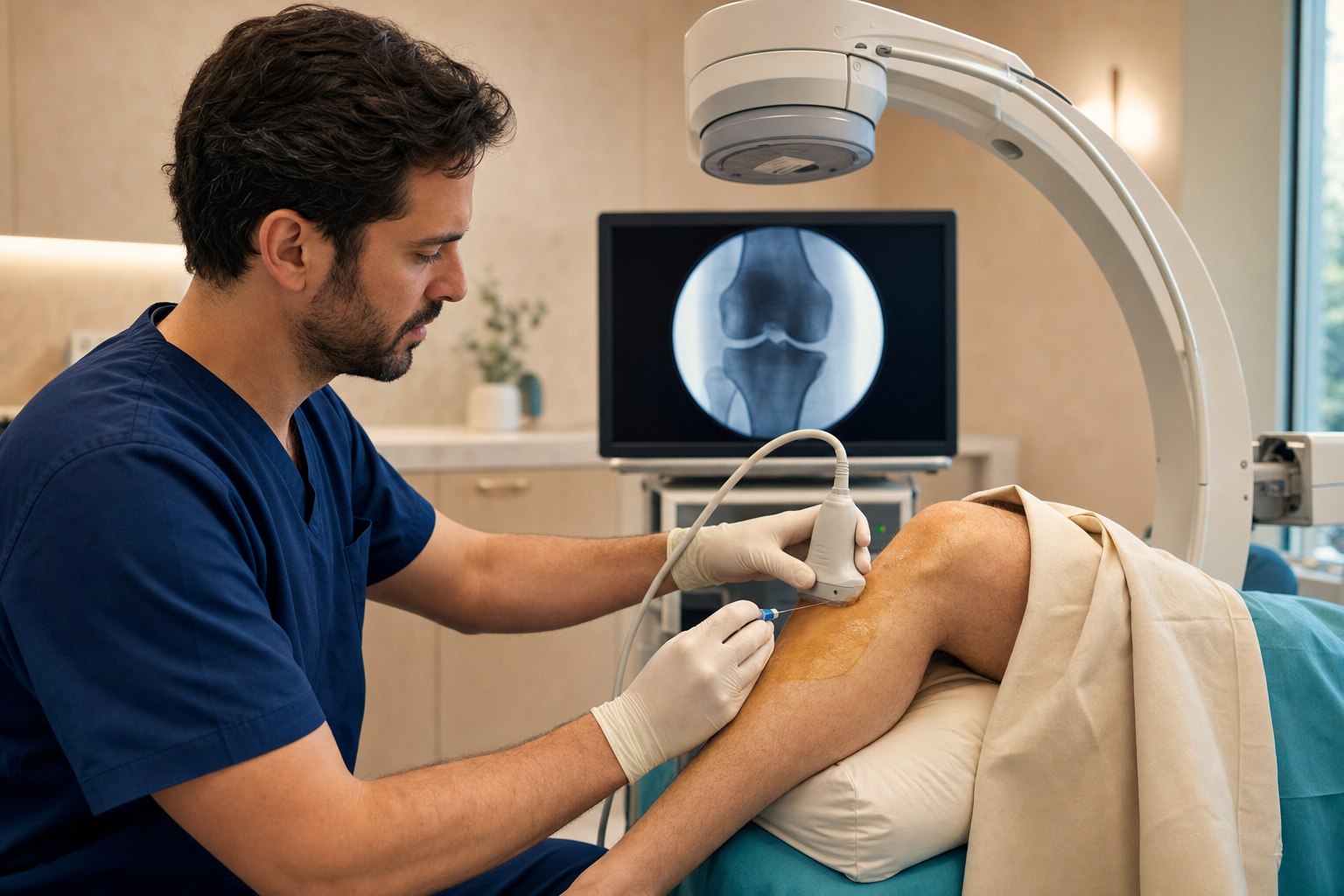
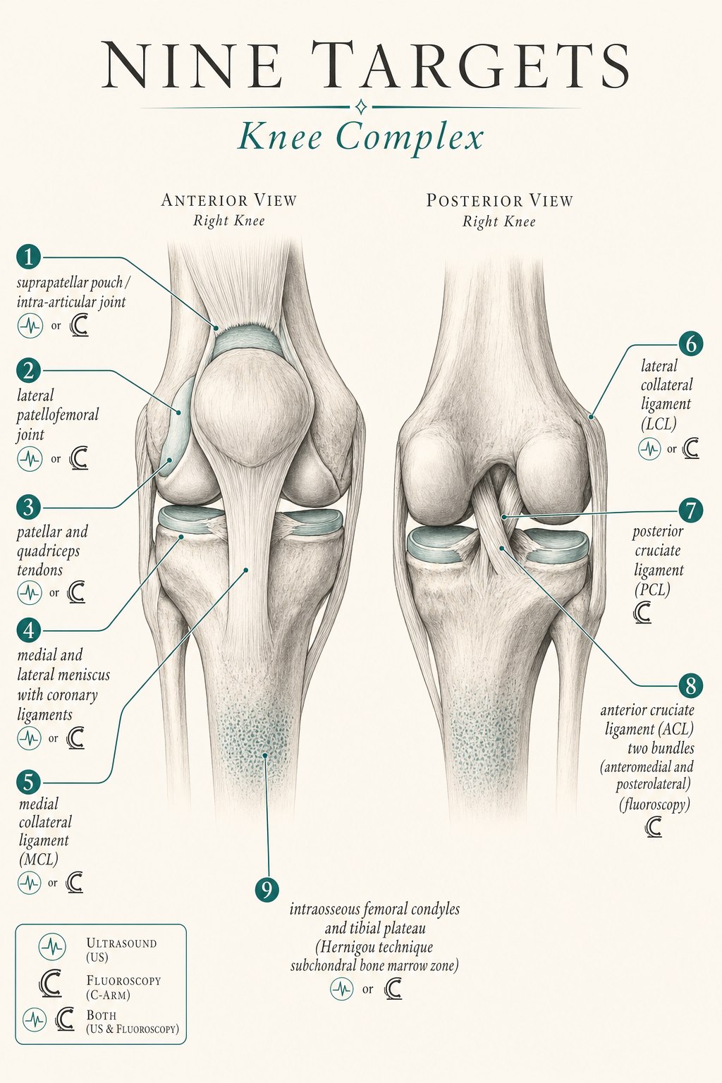
The knee is superficial enough that many injections have historically been done blind, and that habit is one of the reasons a lot of knee patients arrive with a history of injections that “did not work.” Ultrasound is real-time sound imaging — it excels at showing tendon fiber pattern, ligament architecture, meniscal bodies, the synovial pouches of the knee, peripheral nerves, and a needle moving through all of them in real time. For the knee, ultrasound is the imaging tool for the intra-articular joint (via the suprapatellar pouch approach), the lateral patellofemoral joint, the patellar and quadriceps tendons, the meniscal bodies and their coronary suspensory ligaments, the medial and lateral collateral ligaments, and the posterior cruciate ligament. Even at a superficial joint like the knee, the accuracy difference between blind and image-guided injection is meaningful — and at the small collateral and cruciate structures, image guidance is essential rather than optional.
Fluoroscopy is real-time X-ray. In the knee it plays a specific and limited but important role. Two targets require fluoroscopy. The first is the anterior cruciate ligament when we are treating a partial tear (grade 1, grade 2, or a non-retracted grade 3) with an advanced regenerative technique. The ACL has two functional bundles — anteromedial and posterolateral — and a proper biologic treatment of a partial tear requires accurately reaching the femoral and tibial origins and insertions of both bundles. This is the three-needle intraligamentous technique described and refined by Christopher Centeno and the Regenexx group, and it is done under fluoroscopy with a small amount of iodinated contrast to confirm intraligamentous position (Centeno protocol, PMC4527573). The second is the intraosseous compartment of the femoral condyles and tibial plateau — the subchondral bone marrow zone that is the target of the Hernigou technique for advanced osteoarthritis and bone marrow lesions. Bone is invisible to ultrasound; fluoroscopy is the tool that lets the operator place the needle tip accurately in the subchondral bone at the specific location of the pathology.
Here is the safety principle that separates a modern knee procedure from an older one: image guidance is the standard even at a superficial joint. The suprapatellar pouch approach under ultrasound is more accurate than blind injection, avoids the fat pad, and lets the operator confirm that the biologic has diffused into the joint. For the small collateral ligaments, for the meniscal coronary attachments, and for the ACL bundles, image guidance is not a luxury — it is the difference between a real treatment and a hopeful injection.
The nine sections of the knee ensemble
What follows is a section-by-section walk through each structure I target in a comprehensive regenerative knee procedure. For each one I describe what the structure does when it is healthy, how it manifests as pain when it is not, and how it is targeted — including which imaging tool is right for that specific target.
1. The intra-articular joint via the suprapatellar pouch — the main ball and socket of the knee
What it does: The tibiofemoral joint is a modified hinge synovial joint that carries three to five times body weight during walking and up to six times body weight during stair descent. Its articular cartilage is nourished by synovial fluid, and the suprapatellar pouch is the largest anterior extension of the joint cavity, sitting deep to the quadriceps tendon and superior to the patella. It is the safest, most accurate access point for a comprehensive intra-articular knee injection.
How it manifests as pain: Knee osteoarthritis classically presents as anterior or medial joint-line pain, worse with stairs and prolonged standing, morning stiffness that improves within thirty to sixty minutes of activity, crepitus with flexion, loss of full extension and, in advanced disease, loss of full flexion. It can refer to the anterior thigh and the calf. Baker’s cysts and joint effusions are common companions.
How we target it: With an ultrasound-guided suprapatellar pouch intra-articular injection. The evidence base for intra-articular PRP in knee osteoarthritis is now robust and consistent. A systematic review of eighteen Level-I studies found that PRP produced a mean WOMAC improvement of 44.7 percent versus 12.6 percent for hyaluronic acid at twelve months (p<.01) and suggested that leukocyte-poor formulations may be superior (Belk 2020 systematic review, PubMed 32302218). A meta-analysis of fifteen randomized controlled trials with 1,314 patients confirmed that PRP outperforms hyaluronic acid on WOMAC and VAS at six and twelve months (Han 2019 meta-analysis, PubMed 30849177). A 2024 network meta-analysis reported an odds ratio of 2.19 favoring PRP over HA (Xiong 2024 meta-analysis, PubMed 38420745), and emerging data suggest that combined PRP+HA may add a small additional benefit in appropriate patients (Combined PRP+HA meta-analysis, PubMed 39819683). This is one of the most extensively studied applications of regenerative medicine, and image-guided intra-articular PRP for mild-to-moderate knee osteoarthritis is one of the best-supported interventions we offer.
2. The lateral patellofemoral joint — the compartment that quietly runs the front of the knee
What it does: The patellofemoral joint is the articulation between the underside of the patella and the trochlear groove of the femur. It is a compartment in its own right, with its own articular cartilage, its own load pattern, and its own osteoarthritis. It carries roughly three times body weight during a squat and up to seven times body weight during a deep squat or stair descent. Isolated lateral patellofemoral osteoarthritis is common, particularly in patients with a shallow trochlear groove, a lateral patellar tilt, or a history of patellar maltracking or dislocation.
How it manifests as pain: Patellofemoral osteoarthritis presents as anterior knee pain worse with squats, stairs (especially descending), prolonged sitting with the knee bent (the “theater sign”), and crepitus with active knee extension. Tenderness on lateral patellar facet palpation with the knee in extension and a positive patellar grind test are characteristic exam findings. It can be missed on standard AP and lateral X-rays if a skyline (Merchant) view is not obtained.
How we target it: With an ultrasound-guided lateral patellofemoral joint injection. The tibiofemoral compartment does not always communicate freely with the lateral patellofemoral compartment, particularly in patients with adhesive fibrosis or with predominant lateral disease, and a suprapatellar-only injection can miss the exact compartment in pain. Targeting the lateral patellofemoral joint directly under ultrasound ensures the biologic reaches the compartment in question. The evidence base for PRP in patellofemoral osteoarthritis draws from the broader knee osteoarthritis literature and from patellofemoral-specific case series that document reduction in anterior knee pain and improvement in Kujala scores with image-guided PRP.
3. The patellar and quadriceps tendons — the extensor mechanism
What they do: The quadriceps tendon inserts on the superior pole of the patella and transmits the force of the four quadriceps muscles across the knee. The patellar tendon (properly a ligament, since it runs bone to bone) originates on the inferior pole of the patella and inserts on the tibial tuberosity. Together they form the extensor mechanism, which carries the entire extensor load of every squat, every stair climb, every jump, and every landing. Patellar tendinopathy (“jumper’s knee”) is one of the most common overuse injuries in jumping sports; quadriceps tendinopathy is more common in older recreational athletes and in patients with a history of prior extensor mechanism surgery.
How they manifest as pain: Patellar tendinopathy presents as anterior knee pain localized to the inferior pole of the patella, worse with jumping, squatting, and prolonged sitting, tenderness on palpation of the proximal patellar tendon, and pain with a single-leg decline squat. Quadriceps tendinopathy presents as anterior knee pain localized to the superior pole of the patella, worse with resisted knee extension, and tenderness on palpation of the distal quadriceps tendon.
How we target them: With ultrasound-guided PRP into the pathologic tendon body, typically as a series of two to three injections combined with a structured eccentric loading program. I want to be transparent about the state of the evidence here, because it is more mixed than some regenerative marketing suggests. A landmark 2013 trial found that image-guided PRP was superior to focused extracorporeal shock wave therapy at six and twelve months for chronic recalcitrant patellar tendinopathy (Vetrano 2013, PubMed 23408591). A 2025 systematic review and meta-analysis found that PRP was superior to corticosteroid at mid-term follow-up for patellar tendinopathy (2025 meta-analysis, Springer), and a Radiology Advances 2024 network analysis ranked PRP first at 52-week follow-up versus percutaneous tenotomy and sham (Radiology Advances 2024). Balanced against that, a well-designed 2019 randomized trial found that neither leukocyte-rich PRP nor leukocyte-poor PRP was superior to saline when combined with eccentric loading (Scott 2019, PubMed 31038979). Case series using three ultrasound-guided injections combined with eccentric loading have documented return to sport with MRI-demonstrated structural recovery. The honest interpretation: patient selection, injection technique (three-injection protocol into the pathologic tendon body), leukocyte-poor formulation, and rigorous eccentric rehab appear to be the ingredients that separate the successful trials from the negative ones. I discuss the evidence explicitly with every patient before we proceed.
4. The medial and lateral menisci and their coronary suspensory ligaments
What they do: The menisci are two crescent-shaped fibrocartilaginous disks that sit between the femoral condyles and the tibial plateau. They deepen the articular surface, distribute load, absorb shock, provide passive stability, and lubricate the joint. The peripheral third of each meniscus (the “red zone”) has a limited blood supply and can heal; the inner two-thirds (the white zone) is avascular and does not heal on its own. The coronary (or meniscotibial) ligaments are small suspensory ligaments that attach the periphery of each meniscus to the tibia and are a frequent source of joint-line pain when injured.
How they manifest as pain: Meniscal pathology presents as joint-line pain, painful catching or locking, pain with pivoting or twisting, a positive McMurray or Thessaly test on exam, and, in acute injuries, effusion. Degenerative meniscus tears — the horizontal cleavage tears that develop with age and often accompany osteoarthritis — produce a duller, more chronic joint-line pain that is often mistaken for pure osteoarthritis.
How we target them: With ultrasound-guided PRP injection targeting the meniscal body, the joint line, and the coronary ligament attachment. The regenerative evidence for meniscus is meaningfully favorable. A 2018 randomized controlled trial by Kaminski documented an 85 percent healing rate on MRI at eighteen weeks in the PRP-augmented group versus 47 percent in the control group (p=0.048), with sustained improvement in IKDC, WOMAC, and KOOS scores at 42-month follow-up (Kaminski 2018 RCT, PMC5866900). A 2019 percutaneous trephination-plus-PRP study reported a failure rate of 48 percent in the PRP-augmented group versus 70 percent in the control group (p=0.04) (MDPI 2019 trephination + PRP). A 2024 AAOS systematic review found that image-guided PRP for meniscal injury produced MRI improvement in 50.9 percent of patients, significant improvement in Lysholm and KOOS scores, 75 percent return to sport, and 90.7 percent patient satisfaction (AAOS 2024 systematic review). A 2022 meta-analysis by Xie found the picture more mixed at twelve-plus-month follow-up, particularly for degenerative tears in older patients. The technique that appears to work is image-guided PRP delivered to the meniscal body and coronary attachment, ideally for peripheral (red-zone) tears and for degenerative tears in the setting of osteoarthritis. Complex, unstable, displaced meniscal tears with mechanical symptoms of locking remain surgical decisions.
5. The medial collateral ligament (MCL)
What it does: The medial collateral ligament runs from the medial femoral epicondyle to the proximal medial tibia and is the primary restraint against valgus (medial-opening) force at the knee. It has a superficial and a deep portion and blends with the medial meniscus. It is one of the most commonly injured knee ligaments — classically from a lateral blow to the knee in football, from a skiing fall with the foot planted, or from a soccer or basketball cut.
How it manifests as pain: MCL injury presents as medial knee pain and tenderness at the femoral or tibial attachment, pain and laxity with valgus stress at thirty degrees of flexion, bruising or swelling along the medial knee, and, in chronic partial injury, medial-side pain with cutting and pivoting activities. Grade 1 is a stretch injury without laxity; grade 2 is a partial tear with mild laxity; grade 3 is a complete tear with gross laxity.
How we target it: With an ultrasound-guided PRP injection into the specific area of pathology — typically the femoral origin, tibial insertion, or intraligamentous tear. The evidence base is predominantly composed of case reports and case series, but the results in appropriately selected patients are encouraging. A published case documented complete MRI healing of a low-grade MCL injury with ultrasound-guided PRP (Yoshida MCL case, PMC7111026). A 2013 study of an elite footballer with a grade III MCL injury documented return to sport at twenty-five days after PRP-augmented rehabilitation (Eirale 2013, PubMed 23802059). A 2017 three-case series of chronic MCL injuries reported meaningful clinical improvement with image-guided PRP (Bhatia 2017, PubMed 29194097). I am transparent with patients that this is a target where the evidence is emerging rather than mature, and that patient selection — grade 1 and grade 2 partial injuries, non-retracted grade 3 injuries in athletes with clear anatomic pathology and a rehabilitation runway — matters as much as the biologic itself.
6. The lateral collateral ligament (LCL)
What it does: The lateral collateral ligament runs from the lateral femoral epicondyle to the fibular head and is the primary restraint against varus (lateral-opening) force at the knee. It is a component of the posterolateral corner complex, along with the popliteus and popliteofibular ligament, and is less frequently injured in isolation than the MCL. Isolated LCL injuries occur from a medial blow to the knee, from a hyperextension-varus mechanism, and from twisting injuries with the knee in extension.
How it manifests as pain: LCL injury presents as lateral knee pain and tenderness along the ligament, pain and laxity with varus stress at thirty degrees of flexion, and, in chronic injury, lateral-side instability with cutting activities. Because the peroneal nerve wraps around the fibular neck immediately deep to the LCL, LCL injuries are occasionally accompanied by transient or persistent peroneal neuropathy — a critical distinction on exam.
How we target it: With an ultrasound-guided PRP injection into the specific area of pathology, with careful attention to the peroneal nerve immediately deep to the ligament at the fibular head. Ultrasound is essential for LCL work — the peroneal nerve is invisible to fluoroscopy, and a needle placed without visualization near the fibular head carries a real risk of nerve injury. The regenerative evidence for LCL is more limited than for MCL, and case-report level; the technique and indications are extrapolated from the MCL literature and from the broader ligament biologic literature.
7. The posterior cruciate ligament (PCL)
What it does: The posterior cruciate ligament runs from the medial femoral condyle to the posterior tibial plateau and is the primary restraint against posterior tibial translation. It is a shorter, thicker, and biomechanically stronger ligament than the ACL, and its injuries are much less common than ACL injuries — classically resulting from a “dashboard injury” (posteriorly directed force on the flexed knee) or from a fall on a flexed knee with a plantarflexed foot. Isolated grade 1 and grade 2 PCL injuries frequently do well with conservative management; grade 3 PCL injuries and combined ligamentous injuries are typically surgical.
How it manifests as pain: PCL injury presents as poorly-localized posterior or diffuse knee pain, a positive posterior drawer test, a positive sag sign on exam, and, in chronic partial injury, a sense of the knee “giving way” on downhill walking or on stairs. It is one of the most under-diagnosed knee ligament injuries.
How we target it: With a fluoroscopically- or ultrasound-guided PRP injection into the ligament substance. Because the PCL sits behind the anterior structures of the knee, ultrasound access is possible but technically demanding, and many operators use a fluoroscopically-guided posterior approach for grade 1 and grade 2 partial tears in athletes who want to avoid surgery. The evidence base is emerging and consists of small case series; patient selection is critical.
8. The anterior cruciate ligament (ACL) — two bundles, three needles, under fluoroscopy
What it does: The anterior cruciate ligament runs from the posterior aspect of the lateral femoral condyle to the anterior tibial plateau and is the primary restraint against anterior tibial translation and rotational load. It has two functional bundles — the anteromedial bundle (tight in flexion) and the posterolateral bundle (tight in extension) — and the biomechanics of the ligament depend on both bundles being intact. ACL injuries occur in roughly one in three thousand Americans each year, and the majority are non-contact injuries in cutting and pivoting sports. Complete ACL ruptures with retraction are typically surgical in active patients. Partial ACL tears (grade 1, grade 2, and non-retracted grade 3) are a rapidly evolving regenerative target for patients who want to preserve their native ACL and avoid a graft reconstruction.
How it manifests as pain: Acute ACL injury presents as an audible pop, immediate hemarthrosis, pain, and a sense of instability. Partial tears may present with less dramatic acute symptoms and with subsequent instability episodes on cutting and pivoting activities. A positive Lachman test, positive anterior drawer, and positive pivot shift are the physical exam findings; MRI is the imaging standard for staging and for determining whether a tear is complete, partial, or non-retracted.
How we target it: With a fluoroscopically-guided intraligamentous injection of bone marrow aspirate concentrate (BMAC) or leukocyte-poor PRP into the anteromedial and posterolateral bundle origins and insertions — the three-needle two-bundle technique. The technique was described and refined by Christopher Centeno and the Regenexx group and requires fluoroscopy with a small amount of iodinated contrast to confirm intraligamentous position at each of the three needle placements: the femoral origin of both bundles and the tibial insertion. This is an advanced regenerative technique and is appropriate for grade 1 and grade 2 partial tears and for non-retracted grade 3 partial tears in patients who want to avoid reconstruction. Complete tears with retracted stumps remain surgical decisions. The published evidence for intraligamentous BMAC and PRP for partial ACL tears is a growing but still preliminary case-series literature (Centeno protocol, PMC4527573, BiologicOrtho intraligamentous BMAC+PRP). Systematic reviews of PRP as a graft-healing augmentation in surgical ACL reconstruction have been mixed, and the intra-ligamentous injection technique for partial tears — the technique we are describing here — is a distinct application whose evidence base is separate and preliminary but promising. I am transparent about that distinction with every patient we consider for the procedure.
9. The intraosseous femoral condyles and tibial plateau — the Hernigou technique at the knee
What it does: The subchondral bone of the femoral condyles and the tibial plateau sits immediately deep to the articular cartilage of the tibiofemoral joint. It is a metabolically active zone that contains a native population of mesenchymal stem cells, and it is where the earliest changes of advanced osteoarthritis take place. Bone marrow lesions — edema patterns on MRI in the subchondral zone — correlate strongly with pain and progression in knee osteoarthritis and are one of the most reliable radiographic predictors of who will progress toward total knee arthroplasty. The intraosseous injection technique addresses this zone directly, and it is one of the highest-impact developments in modern regenerative orthopedics.
How it manifests as pain: Intraosseous knee pathology presents as a specific pain pattern — deep, poorly-localizable knee pain that is worse with weight-bearing and often worse at rest at night in advanced disease, poorly responsive to intra-articular injection because the pain generator is in the bone rather than in the joint capsule, and correlated on MRI with subchondral bone marrow edema, cystic change, or focal chondral defects. It is the pain pattern that distinguishes patients whose osteoarthritis will respond to an intra-articular injection alone from patients whose disease has progressed to the bone and requires an intraosseous approach.
How we target it: With a fluoroscopically-guided intraosseous injection of bone marrow aspirate concentrate (BMAC) into the femoral condyles and, when appropriate, the tibial plateau — the Hernigou technique applied at the knee. This is the target that most distinguishes a modern regenerative knee practice from a conventional one, and the evidence is genuinely compelling. Hernigou’s 2018 paired-knee comparison — a study of thirty patients with bilateral knee osteoarthritis where one knee received subchondral BMAC and the contralateral knee received total knee arthroplasty — documented comparable Knee Scores at fifteen-year follow-up (78.3 versus 80.3), with 21 of 30 patients preferring the cell-therapy knee. In a broader cohort of 140 patients treated with subchondral BMAC, only 18 required conversion to total knee arthroplasty at a mean of ten years (Hernigou 2018 subchondral BMAC, PubMed 29589086). A 2022 prospective study of combined subchondral and intra-articular BMAC by Boffa documented stable IKDC scores at twenty-four months, statistically significant reduction of bone marrow edema on MRI (p<0.0005), and a 13 percent failure rate (Boffa 2022, PubMed 36326876). A 2024 prospective study of subchondroplasty (a related technique using calcium phosphate rather than BMAC) documented a 76.2 percent arthroplasty-free rate at two years (Subchondroplasty prospective 2024), and a recent meta-analysis of subchondroplasty reported a mean pain VAS reduction of 39 at two years with a 22.4 percent conversion rate to total knee arthroplasty (Subchondroplasty meta-analysis). Two honest caveats. First, the biologic with the evidence is BMAC — a small randomized trial of intraosseous PRP alone (without BMAC) did not add benefit above intra-articular PRP at six months (Intraosseous PRP trial, PubMed 35033356). Second, subchondroplasty (calcium phosphate) and the Hernigou BMAC technique target the same problem with different biologics and different mechanisms; the Hernigou BMAC technique is our preferred approach because the biologic is autologous and provides mesenchymal stem cells rather than a mineral scaffold.
How the conductor puts the knee ensemble together
A comprehensive regenerative knee procedure at Pravida does not treat all nine structures on every patient. The point of the physical exam, the imaging review, and the diagnostic history is to determine which sections of the ensemble are actually out of tune for you. Some patients have a single-structure problem — an isolated patellar tendinopathy in a college volleyball player, an isolated meniscal red-zone tear in a runner, an isolated grade 2 MCL sprain in a skier — and a single well-placed injection is exactly the right answer. Other patients have combined patterns (the most common in adults over fifty is medial-compartment osteoarthritis plus a degenerative medial meniscus tear plus subchondral bone marrow edema plus a compensatory patellar tendinopathy; in cutting-sport athletes it is a partial ACL tear plus a peripheral meniscal tear plus a grade 1 MCL sprain), and treating just one of them is precisely why the last two shots wore off in weeks.
What a world-class knee procedure looks like on the day it happens: a physical exam and imaging review (including a weight-bearing X-ray series with a skyline patellofemoral view and a recent MRI when appropriate) that identifies the specific structures in play, a written plan for which structures will be treated, a procedure suite set up with both a high-resolution ultrasound machine and a C-arm fluoroscope, and a sequence of small, precise injections — each one confirmed on real-time imaging before medication is delivered — that treats each identified section of the ensemble with the tool that shows it best. Ultrasound is used for the intra-articular joint via the suprapatellar pouch, the lateral patellofemoral joint, the patellar and quadriceps tendons, the meniscal bodies and coronary ligaments, the medial and lateral collateral ligaments, and often the PCL. Fluoroscopy is used for the ACL three-needle two-bundle technique and for the intraosseous Hernigou work at the femoral condyles and tibial plateau. Most knee procedures are outpatient and take under ninety minutes. Most patients drive themselves home the same day, though weight-bearing precautions apply to the intraosseous protocol and to grade 2-3 ligament work.
The choice of biologic — platelet-rich plasma (typically leukocyte-poor for tendon, ligament, and meniscal work), bone marrow concentrate for higher-grade partial ligament injury, advanced joint disease, ACL bundle work, or intraosseous work, or five percent dextrose for adjunctive nerve hydrodissection at the pes anserinus — is guided by the tissue being treated and by the patient’s overall regenerative plan. The CartiNova program is our organized framework for making those choices in a way that is transparent, evidence-informed, and personalized to the individual patient.
What most knee patients get wrong before they see us
- They accept a blind knee shot as the standard first step. The suprapatellar pouch approach under ultrasound is more accurate than blind injection, avoids the fat pad, and lets the operator confirm that the biologic has diffused into the joint. Image-guided technique is the modern standard even at a superficial joint.
- They treat one compartment and ignore the others. Anterior knee pain from patellofemoral disease frequently coexists with medial-compartment osteoarthritis, and a single suprapatellar-only injection often misses the exact compartment in pain. A comprehensive evaluation identifies which of the nine structures are actually generating symptoms.
- They ignore the bone. In advanced osteoarthritis, the intra-articular injection often wears off in weeks because the pain generator is in the subchondral bone, not in the joint capsule. Bone marrow lesions on MRI are the specific indication for the Hernigou intraosseous BMAC technique — the intervention that changes the disease trajectory when it is done correctly and with the right biologic.
- They confuse an injection decision with a surgical decision. Complete retracted ACL rupture in a cutting-sport athlete is a reconstruction decision. Complete unstable displaced meniscal tears with locking are meniscectomy or repair decisions. Bone-on-bone stage 4 osteoarthritis with severe deformity is a total knee arthroplasty decision. A good regenerative practice will tell you clearly when injection is the right answer and when it is not.
- They expect regeneration without rehabilitation. A well-placed biologic creates a window in which targeted knee strengthening, glute and hip strengthening, and progressive loading exercises can rebuild the tissue and restore function. The injection and the rehab are one intervention, not two.
The honest limits, and where a good clinician still matters
- The nine-structure approach is designed for patients with chronic or subacute knee pain that has not fully resolved with conservative care. It is not first-line treatment for advanced (stage 4) knee osteoarthritis with severe deformity, complete retracted ACL rupture in a cutting-sport athlete who wants to return to competitive sport, complete unstable displaced meniscal tears with locking, complete grade 3 collateral rupture with functional instability in a young athlete, acute traumatic knee dislocation, septic arthritis, or red-flag presentations (fever, unexplained weight loss, night pain unresponsive to positional change, or pain accompanied by new neurologic deficit).
- The regenerative evidence base is strongest for image-guided intra-articular PRP in knee osteoarthritis, for the Hernigou intraosseous BMAC technique in advanced osteoarthritis with bone marrow lesions, and for image-guided PRP for peripheral (red-zone) and degenerative meniscal tears. It is meaningful and developing for patellar tendinopathy (with best results using leukocyte-poor PRP, three-injection protocols, and structured eccentric loading). It is emerging and case-series level for MCL, LCL, PCL, and partial ACL tears — where patient selection, technical precision, and rehabilitation are as important as the biologic itself. I discuss the strength of the evidence for the specific target with every patient before we proceed.
- Any advanced ACL biologic technique requires fluoroscopy. The three-needle two-bundle intraligamentous injection cannot be performed accurately under ultrasound alone. A clinician who offers “ACL injection” without fluoroscopic guidance is not performing the technique that the published protocol describes.
- Any LCL work requires ultrasound because of the peroneal nerve. A fluoroscopically-guided-only injection near the fibular head risks peroneal nerve injury.
- Regenerative outcomes depend on the underlying tissue, the biologic used, the technical precision of delivery, and the rehabilitation that follows. A biologic delivered blindly, or into the wrong structure, is not regenerative — it is just an injection.
- If you have new onset of severe knee pain with fever, sudden inability to bear weight after minor trauma, pain accompanied by new neurologic deficit, or new knee pain in the setting of active cancer, please contact your physician or an emergency department today. Those are not situations for an elective regenerative procedure.
Ready to have your specific knee pattern mapped?
If you are an Atlanta-area patient with chronic knee pain that has not fully responded to prior injections or conservative care — or you are researching alternatives to total knee arthroplasty, ACL reconstruction, or meniscectomy before you commit to surgery — we would rather see you in person. A consultation at Pravida Health includes a physical exam, an imaging review, and, where appropriate, a comprehensive regenerative plan tailored to the specific sections of your knee ensemble that are out of tune.
Book a consultationKey sources referenced in this article
- Belk JW, et al. PRP versus hyaluronic acid for knee osteoarthritis: systematic review of eighteen Level-I studies. PubMed 32302218. PRP produced 44.7 percent WOMAC improvement versus 12.6 percent HA at twelve months (p<.01); leukocyte-poor PRP may be superior.
- Han Y, et al. PRP versus hyaluronic acid for knee osteoarthritis: meta-analysis of fifteen RCTs with 1,314 patients. PubMed 30849177. PRP superior on WOMAC and VAS at six and twelve months.
- Xiong Y, et al. Network meta-analysis of PRP versus HA for knee osteoarthritis, 2024. PubMed 38420745. Odds ratio 2.19 favoring PRP.
- Combined PRP+HA meta-analysis for knee osteoarthritis. PubMed 39819683. Combination therapy may add small benefit in appropriate patients.
- Vetrano M, et al. PRP versus ESWT for chronic patellar tendinopathy: randomized controlled trial. PubMed 23408591. Image-guided PRP superior at six and twelve months.
- Scott A, et al. LR-PRP versus LP-PRP versus saline with eccentric loading for patellar tendinopathy. PubMed 31038979. Neither PRP formulation superior to saline when combined with eccentric loading — important negative RCT.
- Systematic review and meta-analysis of PRP versus corticosteroid for patellar tendinopathy, 2025. Springer 2025 meta-analysis. PRP superior to corticosteroid at mid-term follow-up.
- Radiology Advances 2024 network analysis for patellar tendinopathy treatments. Radiology Advances 2024. PRP ranked first at 52-week follow-up versus percutaneous tenotomy and sham.
- Kaminski R, et al. PRP-augmented meniscal repair: randomized controlled trial. PMC5866900. 85 percent healing at 18 weeks versus 47 percent (p=0.048); sustained IKDC, WOMAC, KOOS improvement at 42 months.
- Percutaneous trephination plus PRP for meniscal tears, 2019. MDPI 2019. Failure rate 48 percent PRP versus 70 percent control (p=0.04).
- AAOS 2024 systematic review of image-guided PRP for meniscal injury. AAOS 2024. 50.9 percent MRI improvement, significant Lysholm and KOOS improvement, 75 percent return to sport, 90.7 percent satisfaction.
- Yoshida M, et al. Complete MRI healing of low-grade MCL injury after ultrasound-guided PRP: case report. PMC7111026.
- Eirale C, et al. Grade III MCL injury in an elite footballer: return to sport at 25 days with PRP. PubMed 23802059.
- Bhatia N, et al. Chronic MCL injury treated with image-guided PRP: three-case series. PubMed 29194097.
- Centeno CJ, et al. Intraligamentous BMAC and PRP for partial ACL tears: three-needle two-bundle technique under fluoroscopy. PMC4527573. Technical description and preliminary outcomes.
- BiologicOrtho 2023 intraligamentous BMAC+PRP for partial ACL. BiologicOrtho, 2023.
- Hernigou P, et al. Subchondral BMAC for knee osteoarthritis: paired-knee comparison against contralateral total knee arthroplasty at 15 years. PubMed 29589086. Comparable Knee Scores; 21 of 30 patients preferred cell-therapy knee; only 18 of 140 patients required TKA at ten years.
- Boffa A, et al. Combined subchondral and intra-articular BMAC for knee osteoarthritis: 24-month prospective. PubMed 36326876. Stable IKDC; significant reduction in bone marrow edema (p<0.0005); 13 percent failure rate.
- Subchondroplasty prospective outcomes 2024. PubMed 39667406. 76.2 percent arthroplasty-free at two years.
- Subchondroplasty meta-analysis. PubMed 41950892. Mean VAS reduction 39 at two years; 22.4 percent TKA conversion.
- Intraosseous PRP alone versus intra-articular PRP alone for knee osteoarthritis: randomized trial. PubMed 35033356. Intraosseous PRP did not add benefit at six months — BMAC, not PRP, is the biologic with the intraosseous evidence.