
The wrist and hand as a precision string quartet
The wrist and hand are the most tightly packed piece of the musculoskeletal system in the body. Twenty-seven bones, more than thirty joints, thirty-four muscles firing through a tunnel narrower than a wedding ring, three major nerves and dozens of digital branches, and a set of ligaments that together allow both the delicate rotation of a violinist’s bow and the crushing grip of a rock climber — all in a space smaller than a paperback book. When any one of those pieces is out of tune, the rest of the ensemble strains to compensate, and the pain rarely stays where the actual injury lives.
This is the fourth article in a series on how a master regenerative procedure actually works, region by region. We started with the five-structure ensemble of the lumbar spine, walked through the fourteen-structure symphony of the shoulder, and mapped the seven-structure ensemble of the elbow. The hand and wrist require a different mindset than any of those. There is essentially no redundancy here — a millimeter separates a therapeutic injection from a nerve injury, and the classic “steroid shot for the wrist” is almost never precise enough to be the right first choice. Precision matters more here than anywhere else in the upper limb, and precision depends on imaging.
What follows is the conductor’s map for the hand and wrist: every structure I target in a comprehensive regenerative wrist and hand procedure, why each one matters, how it produces pain when it is out of tune, and which imaging tool — ultrasound, fluoroscopy, or both — is right for that section.
Why two imaging tools, and why ultrasound is the dominant one in the hand
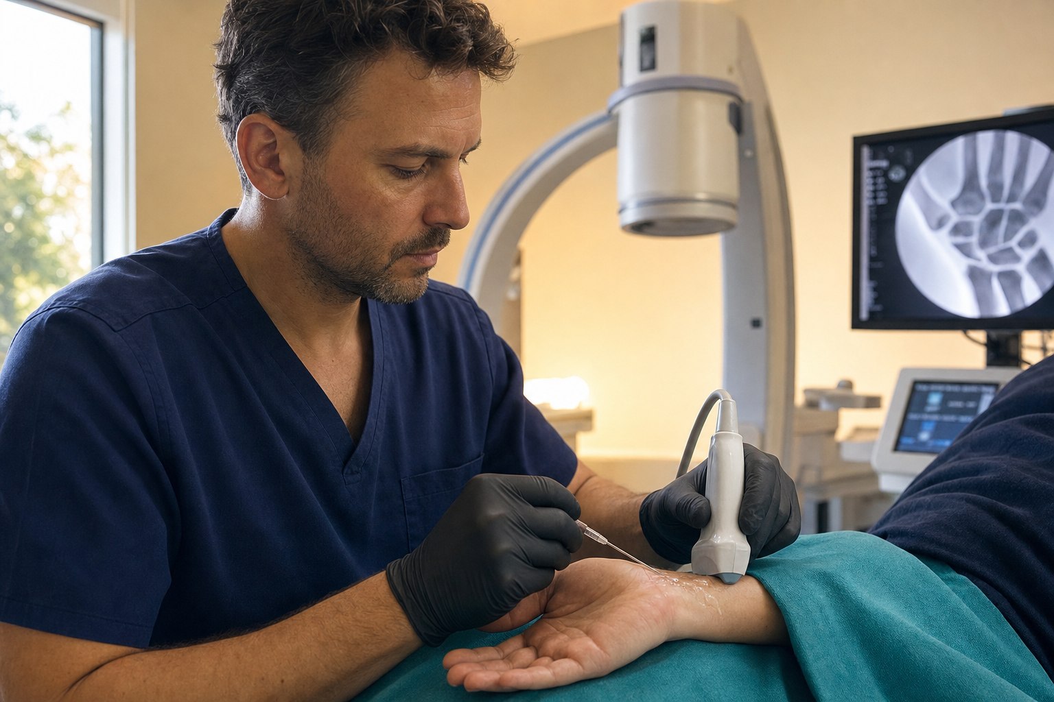
The same imaging philosophy we applied to the elbow applies here, but with the balance tipped further toward ultrasound. Ultrasound is real-time sound imaging — it excels at showing tendon fibers, ligament architecture, capsular thickness, and, most importantly, peripheral nerves and a needle moving through all of them in real time. In the hand and wrist, ultrasound is the imaging tool. Cadaveric studies of the thumb CMC joint show ultrasound-guided injection accuracy near 94 percent versus roughly 70 percent for landmark-based technique (Umphrey et al., cadaver accuracy study), and clinical outcome studies show meaningful superiority of image-guided over palpation-guided technique for exactly this reason (Frontiers in Pharmacology, network review 2021).
Fluoroscopy is real-time X-ray. In the wrist it plays a targeted, supporting role: intra-articular confirmation of the thumb CMC or the radiocarpal joint when the joint is severely degenerated and capsular constraint makes ultrasound-only entry uncertain, and, in select cases, arthrographic mapping of scapholunate integrity when we want to see whether dye flows through a suspected ligament defect.
Here is the safety caveat that separates a safe wrist procedure from an unsafe one: the peripheral nerves of the wrist and hand are invisible to fluoroscopy. The median nerve at the carpal tunnel, the radial nerve superficial branch as it crosses the first extensor compartment (right where De Quervain patients hurt), the ulnar nerve at Guyon canal, and the digital nerves alongside every flexor tendon sheath are all critical structures that fluoroscopy simply cannot show. For any injection at the volar wrist, the radial side of the wrist near De Quervain territory, or the finger flexor sheaths, ultrasound is not optional. It is the standard.
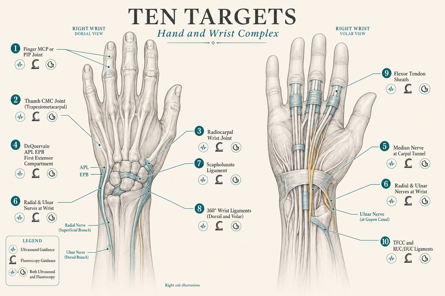
The ten sections of the wrist and hand ensemble
What follows is a section-by-section walk through each structure I target in a comprehensive regenerative wrist and hand procedure. For each one I describe what the structure does when it is healthy, how it manifests as pain when it is not, and how it is targeted — including which imaging tool is right for that specific target.
1. The finger joints (MCP and PIP) — small joints, large functional impact
What they do: The metacarpophalangeal (MCP) joints are the knuckles at the base of the fingers; the proximal interphalangeal (PIP) joints are the middle knuckles. Together with the distal interphalangeal (DIP) joints, they give the human hand its uniquely dexterous range. Each is a tightly capsulated joint supported by collateral ligaments, a volar plate, and the extensor apparatus. They are the joints most affected by osteoarthritis in the hand, and the PIPs in particular carry a disproportionate burden of post-traumatic arthritis in climbers, ball-sport athletes, and manual workers.
How they manifest as pain: MCP and PIP arthritis present as deep aching at the joint, stiffness worst in the morning, loss of full flexion (a difficulty making a tight fist), pain with gripping, and, over time, visible nodularity and deformity. Inflammatory arthritides (rheumatoid, psoriatic) target these joints early and require systemic treatment; osteoarthritis of the PIPs is the most common non-inflammatory presentation and is where a well-placed regenerative injection can meaningfully change the arc of the disease.
How we target them: With an ultrasound-guided intra-articular injection into the specific finger joint. These joints are small — often three to four millimeters wide — and their capsules are close to the extensor tendon, the collateral ligaments, and the digital nerves. Blind injection has a high miss rate and a real risk of injuring adjacent structures; ultrasound reliably confirms intra-articular placement in a target the width of a matchhead. The specific biologic (PRP or, for advanced disease with subchondral involvement, bone marrow concentrate) is chosen based on the stage of disease and the tissue being treated.
2. The thumb carpometacarpal (CMC) joint — the most-worked joint in the hand
What it does: The thumb CMC (or trapeziometacarpal) joint is the base of the thumb, where the first metacarpal meets the trapezium. It is a saddle joint that gives the thumb its unique opposable range — the motion that lets us pinch, grip, and grasp. The CMC is the second-most-common site of osteoarthritis in the entire body, second only to the knee, and it is the number-one cause of chronic hand pain in adults over fifty. Cortisone shots at this joint are one of the most common orthopedic injections in the United States.
How it manifests as pain: CMC arthritis presents as pain at the base of the thumb worse with pinch (turning a key, opening a jar, twisting a doorknob), tenderness right over the joint, a positive grind test, and, in more advanced cases, visible squaring of the joint and progressive adduction contracture of the thumb. It is a joint where meaningful function — the strength of the pinch that lets us open a childproof medication bottle — disappears years before radiographic disease looks “bad enough” for a surgeon.
How we target it: With ultrasound-guided intra-articular injection at the thumb CMC, with fluoroscopic arthrographic confirmation added when capsular constraint or advanced disease makes ultrasound-only entry uncertain. This is one of the marquee applications of orthobiologic medicine. A randomized controlled trial comparing PRP with corticosteroid at 12 months found PRP superior on VAS pain (p=0.015), DASH function score (p=0.025), and patient satisfaction (70 percent versus 13 percent, p=0.002) — a durable, clinically meaningful advantage (Malahias et al., 2018 RCT). A long-term case series of ultrasound-guided PRP for CMC arthritis found 68.8 percent of patients reported improvement with a mean duration of symptom benefit of 15.6 months from a single injection series (Long-term PRP outcomes). A safety and satisfaction review of 43 patients found 73.7 percent patient satisfaction with no significant complications (Safety and satisfaction review, 2025). Ultrasound alone is accurate 94 percent of the time in cadaveric study, and 25 percent more accurate than blind injection in clinical practice (Umphrey cadaver study). This is the joint where the difference between a landmark-guided injection and an image-guided one is the difference between an unpredictable result and a reproducible one.
3. The radiocarpal wrist joint — the deep hinge of the wrist
What it does: The radiocarpal joint is the true wrist joint — the articulation between the distal radius, the proximal row of carpal bones (scaphoid, lunate, triquetrum), and the triangular fibrocartilage complex that supports the ulnar side. Every wrist flexion, extension, radial and ulnar deviation motion happens here. It is the joint that becomes arthritic after distal radius fracture, after scapholunate ligament failure in the SLAC wrist pattern, and in inflammatory arthritis of the wrist.
How it manifests as pain: Radiocarpal joint pain presents as deep dorsal wrist pain, worse with end-range flexion and extension, stiffness, loss of terminal range, and, in advanced cases, a visible bump at the dorsal wrist from dorsal osteophyte formation. It is often accompanied by pain with any forceful grip because grip loading transmits force through this joint.
How we target it: With an intra-articular radiocarpal injection under ultrasound, with fluoroscopic arthrography added when the joint is severely constrained or when arthrographic mapping of a suspected ligament tear is desired. Ultrasound-guided dorsal approaches into the radiocarpal joint are highly accurate in trained hands. When we want to confirm that a communicating tear allows fluid to flow between the radiocarpal, midcarpal, and DRUJ compartments — a diagnostic question — we add fluoroscopy with a small amount of iodinated contrast. This is one of the wrist targets where both imaging tools are used together, and using both is what turns a good injection into a diagnostic and therapeutic one.
4. The first extensor compartment — De Quervain tenosynovitis
What it does: The first dorsal extensor compartment of the wrist contains two tendons — the abductor pollicis longus (APL) and the extensor pollicis brevis (EPB) — that share a single tight fibrous tunnel over the radial styloid on their way to the thumb. Every time we lift a child under the arms, wring out a towel, or make a repeated radial-deviation motion at the wrist, these two tendons glide through that tunnel. Chronic overload causes the tendon sheath to thicken, and glide is progressively lost. Adjacent to the compartment is the intersection with the second compartment (extensor carpi radialis longus and brevis) — the site of the related but distinct intersection syndrome.
How it manifests as pain: De Quervain tenosynovitis presents as radial-side wrist pain right over the radial styloid, worse with thumb use and with ulnar deviation of the wrist, and a positive Finkelstein or Eichhoff test. Intersection syndrome presents as pain and swelling a few centimeters proximal to the wrist — roughly four to eight centimeters proximal to the radial styloid — often with a palpable crepitus described as squeaking or crunching. It is a favorite condition of rowers, cross-country skiers, and any occupation involving repetitive wrist extension against resistance.
How we target it: With ultrasound-guided injection into the first extensor compartment or, for intersection syndrome, into the second extensor compartment or the intersection itself. Two important safety and evidence points define this target. First, the superficial radial nerve passes across the first extensor compartment; a corticosteroid injection performed blindly at this site risks direct nerve injury, subcutaneous fat atrophy, and skin depigmentation — complications that have been reported in roughly 27 percent of blind corticosteroid injections but essentially 0 percent of image-guided PRP injections in randomized comparison (Kumar et al., Journal of Wrist Surgery, 2022). Second, and more importantly, the durability of PRP versus corticosteroid diverges at longer follow-up. A randomized trial found both effective in the short term but with a shift toward PRP superiority by 6 months (Sagar et al., 2024 RCT). A 2024 systematic review of PRP versus corticosteroid for De Quervain found PRP significantly superior at 6 months on VAS pain (p<0.001) with fewer complications (p=0.026) (Systematic review and meta-analysis, 2025). A 2024 RCT confirmed the pattern: corticosteroid superior at 2 weeks, PRP superior at 6 months (Kim et al., RCT 2024). Ultrasound-guided hydrodissection of the first compartment with dextrose is another option, particularly for patients who wish to avoid steroid entirely (D5W hydrodissection technique). For intersection syndrome, an image-guided injection into the second compartment or the intersection is the intervention of choice (Ultrasound technique for intersection syndrome). Ultrasound is essential for both.
5. The median nerve at the carpal tunnel — hydrodissection for carpal tunnel syndrome
What it does: The median nerve is the main nerve of the front of the wrist and hand. It passes through the carpal tunnel — the fibro-osseous channel formed by the carpal bones and the transverse carpal ligament — on its way to supplying sensation to the thumb, index, middle, and half of the ring finger, and motor control of the thenar muscles that let us oppose the thumb. Carpal tunnel syndrome is the most common compression neuropathy in the human body.
How it manifests as pain: Carpal tunnel syndrome presents as numbness and tingling in the thumb, index, middle, and radial half of the ring finger, worse at night when patients wake with the hand asleep, worse with prolonged wrist flexion (driving, phone use, typing), progressive weakness of thumb opposition, and, in advanced cases, thenar atrophy that is visible to the naked eye. It is one of the most common conditions in adult primary care, and it is one of the conditions where the standard-of-care intervention has quietly shifted over the past decade — from cortisone or surgical release toward ultrasound-guided nerve hydrodissection.
How we target it: With ultrasound-guided perineural hydrodissection of the median nerve at the carpal tunnel using five percent dextrose in water (D5W) and, in select cases, PRP. Hydrodissection means using a fluid to gently separate the nerve from the surrounding transverse carpal ligament and flexor tendons that are compressing it. It restores the natural gliding motion of the nerve and, in the process, decompresses the nerve without surgery. The evidence base for this technique has become remarkably robust. A 2024 network meta-analysis of injectable treatments for carpal tunnel syndrome ranked five percent dextrose as most effective at 12 weeks and PRP as most effective at 24 weeks, with a SUCRA of 95.7 for PRP at long-term follow-up (Yonsei Medical Journal network meta-analysis, 2024). A randomized trial identified 10 mL as the optimal D5W hydrodissection volume (Optimal D5W volume RCT). PRP has shown superiority over corticosteroid at long-term follow-up while producing equivalent short-term relief (PRP vs corticosteroid RCT, Long-term follow-up trial). In patients with persistent symptoms after carpal tunnel release surgery, D5W hydrodissection produced effective outcomes in 61 percent of patients with 33-month mean symptom durability (Post-release CTS hydrodissection series) — a meaningful salvage option for patients whose surgery did not fix the problem. Ultrasound is essential; the median nerve is invisible to fluoroscopy.
6. The radial and ulnar nerves at the wrist — regional anesthesia and entrapment
What they do: The radial nerve superficial branch supplies sensation to the dorsal thumb web and radial side of the hand; the ulnar nerve at the wrist (through Guyon canal) supplies sensation to the little finger, half of the ring finger, and motor control of the intrinsic muscles of the hand. Both nerves can be blocked at the wrist for regional anesthesia during hand procedures, and both can be sites of entrapment neuropathy — radial nerve entrapment at the first extensor compartment (Wartenberg syndrome), ulnar nerve entrapment at Guyon canal (typically from repetitive handlebar pressure in cyclists, or from a hook of hamate fracture).
How they manifest as pain: Radial nerve entrapment at the wrist presents as dorsoradial hand pain and numbness worse with tight wristwatch or handcuff use. Ulnar nerve entrapment at the wrist presents as little-finger and ring-finger numbness with weakness of grip, distinguishable from cubital tunnel by sparing of the dorsal ulnar hand sensation (which the dorsal ulnar cutaneous branch supplies proximal to Guyon canal).
How we target them: With ultrasound-guided perineural block for regional anesthesia, or ultrasound-guided hydrodissection with D5W or platelet lysate for entrapment neuropathies. Ultrasound-guided perineural blocks with D5W hydrodissection have been shown non-inferior to standard local anesthetic blocks for wrist and hand regional anesthesia (D5W perineural block non-inferiority), and ultrasound-guided D5W has been shown to produce over 88 percent analgesia rates in wrist and hand nerve blocks (Wiley 2017 outcome study). For entrapment neuropathies, the same hydrodissection principles that apply to the median nerve at the carpal tunnel apply to the radial and ulnar nerves at the wrist. Ultrasound is again essential.
7. The scapholunate ligament — the keystone of carpal stability
What it does: The scapholunate ligament connects the scaphoid and lunate carpal bones on the dorsal side of the wrist. It is the single most important intrinsic wrist ligament — the keystone of proximal-row carpal kinematics. When it is intact, the scaphoid and lunate move as one under load, and the wrist absorbs force efficiently. When it is disrupted, the scaphoid flexes and the lunate extends, creating dorsal intercalated segment instability (DISI), which over years progresses to a specific pattern of degenerative arthritis called SLAC wrist — the most common form of wrist arthritis.
How it manifests as pain: Scapholunate injury presents as dorsal wrist pain worse with loaded extension (a push-up, a fall on outstretched hand, pushing off a chair to stand), pain with a positive scaphoid shift (Watson) test, and, in later stages, a visible clunk on radial-to-ulnar deviation loading. Staging matters: predynamic and dynamic instability (stages 1 and 2) are the window in which regenerative treatment can potentially alter the disease trajectory. Static instability with DISI on plain films (stage 3) and SLAC arthritis (stage 4) are surgical decisions.
How we target it: With ultrasound-guided injection into or along the scapholunate ligament, sometimes with fluoroscopic arthrographic confirmation of the extent of the tear. This is a target where I want to be honest about the state of the evidence. The regenerative literature for scapholunate ligament injury is limited and largely emerging — case series and single-center reports rather than randomized trials, and a recent scoping review noted “no specific data supporting biologic use in carpal instability” as a mature body of evidence (PM&R scoping review, 2023). Case series in predynamic and dynamic instability suggest that image-guided orthobiologic injection at the scapholunate ligament may improve stability and reduce pain at six months in appropriately selected stage 1-2 patients, but the evidence is not yet where it is for the CMC joint or the carpal tunnel. What I offer patients is transparent: for stage 1-2 scapholunate injury this is a legitimate emerging option; for stage 3-4 disease this is a surgical decision and biologic therapy is not the right answer. The imaging tool is ultrasound with occasional fluoroscopic support for arthrographic mapping.
8. The 360-degree wrist ligaments — dorsal and volar carpal stability
What they do: The intrinsic and extrinsic ligaments of the wrist wrap the carpal bones on both the dorsal and volar sides. On the dorsal side, the dorsal radiocarpal, dorsal intercarpal, and radiolunotriquetral ligaments provide extension and rotational stability. On the volar side, the radioscaphocapitate, long and short radiolunate, ulnolunate, and ulnotriquetral ligaments — the “V ligaments” — provide flexion stability and prevent ulnar translation of the carpus. Together they are what allows the wrist to bear a bench-press-worthy load without dissolving into instability.
How they manifest as pain: Chronic wrist instability after ligament injury presents as vague, poorly-localized wrist pain worse with loading, a sensation of “giving way,” positional discomfort at end range, and progressive weakness of grip because the wrist can no longer transmit force from forearm to hand efficiently. It is common after a fall on outstretched hand that was “just a sprain” on the initial X-ray, and it is common in gymnasts, climbers, and manual workers with a history of accumulated microtrauma.
How we target them: With ultrasound-guided injection of specific dorsal or volar ligaments identified as pathologic on imaging, treating the wrist as a 360-degree structure. The technical philosophy borrowed from the pioneering work in interventional regenerative orthopedics is that the wrist ligaments must be treated as a system, not as isolated points — if one restraint is compromised, others are likely overloaded, and comprehensive assessment of both dorsal and volar bands is what defines a well-designed procedure. Evidence here is largely from case series and expert consensus rather than randomized trials, and I set expectations accordingly. Ultrasound is essential; these ligaments are invisible to fluoroscopy.
9. The flexor tendon sheath — trigger finger and flexor tenosynovitis
What it does: The flexor tendon sheath is the tight fibrous tunnel that guides each finger’s flexor tendon from the palm to the fingertip. The A1 pulley at the base of each finger is the most commonly involved site of trigger finger — when the tendon nodularity or sheath thickening exceeds a critical threshold, the tendon catches, locks, and then pops through the pulley with a painful snap. It is one of the most common hand conditions in patients with diabetes and in patients over fifty.
How it manifests as pain: Trigger finger presents as pain and tenderness at the base of the affected finger on the volar side, a palpable nodule that moves with tendon glide, catching or locking on flexion or extension, and, in advanced cases, a finger locked in flexion that requires the other hand to pry open. Flexor tenosynovitis without triggering presents as diffuse volar hand or finger pain worse with grip.
How we target it: With ultrasound-guided injection into the flexor tendon sheath at the A1 pulley, with hydrodilatation of the sheath itself in selected cases. Ultrasound guidance ensures the injection is intrasheath rather than intratendinous — a distinction that matters because intratendinous injection risks tendon damage. Hydrodilatation with volume expansion of the sheath — a variant of hydrodissection technique — can mechanically release adhesions and improve tendon glide. Ultrasound is essential; the digital nerves run immediately adjacent to the flexor sheath and blind injection carries real risk.
10. The triangular fibrocartilage complex (TFCC) and thumb radial collateral ligament
What it does: The TFCC is the cartilage-and-ligament complex on the ulnar side of the wrist — the structure that supports the ulnocarpal joint and stabilizes the distal radioulnar joint (DRUJ). Its central disc is fibrocartilage; its peripheral ligaments (radioulnar collateral, dorsal ulnocarpal, palmar ulnocarpal, and the meniscal homologue) provide the stability that lets the forearm rotate freely under load. TFCC tears are a leading cause of ulnar-sided wrist pain and are especially common in racquet-sport athletes, gymnasts, and patients with a history of fall on outstretched hand with wrist extension and ulnar deviation.
How it manifests as pain: TFCC injury presents as ulnar-sided wrist pain worse with pronation-supination under load, a positive fovea sign (tenderness in the soft spot between the ulnar styloid and the FCU tendon), and, in more severe cases, DRUJ instability. Class 1 (traumatic) tears involve the disc; Class 2 (degenerative) tears involve the disc and often adjacent chondromalacia. The thumb radial collateral ligament (of the MCP joint) is a distinct but related target — injury from forced ulnar deviation of the thumb (a “skier’s thumb”) that classically involves the ulnar collateral ligament also involves the radial side in a smaller subset of patients, and chronic laxity of the RCL of the thumb MCP is a legitimate regenerative target.
How we target them: With ultrasound-guided injection into the TFCC or the specific ulnar-sided ligament (RUC, DUC) implicated on imaging, and with ultrasound-guided injection at the thumb MCP radial collateral ligament for chronic laxity. The TFCC evidence base has developed meaningfully in the past several years. A prospective case series of ultrasound-guided PRP for TFCC tears documented PRWE scores falling from 55.83 to 26.67 at 8 weeks (p=0.003) with sustained benefit through the follow-up period (Thieme prospective series). A case report of leukocyte-poor PRP for TFCC tear documented 70 percent pain relief at 7 months and complete pain resolution at 9 months (LP-PRP case report). A six-patient case series from the Hong Kong Institute of Musculoskeletal Medicine documented VAS scores falling from 5.67 to 0.83 with three of six patients reporting greater than 80 percent relief (HKIMM series). An important caveat: in patients who are already surgical candidates for TFCC repair, adding PRP does not improve the outcome above the surgical repair itself (TFCC repair with vs. without PRP RCT) — injection is the intervention for the patient in whom surgery is not the right choice, not an add-on for a surgical patient. Ultrasound is essential for both TFCC and thumb RCL work.
How the conductor puts the wrist and hand ensemble together
A comprehensive regenerative wrist and hand procedure at Pravida does not treat all ten structures on every patient. The point of the physical exam, the imaging review, and the diagnostic history is to determine which sections of the ensemble are actually out of tune for you. Some patients have a single-structure problem — an isolated CMC arthritis, an isolated trigger finger, an isolated median nerve at the carpal tunnel — and a single well-placed injection is exactly the right answer. Other patients have combined patterns (the most common in adults over fifty is CMC arthritis plus median nerve compression; in racquet-sport athletes it is TFCC plus radiocarpal joint synovitis; in cyclists it is ulnar nerve at Guyon canal plus wrist ligament irritation), and treating just one of them is precisely why the last two shots wore off in weeks.
What a world-class wrist and hand procedure looks like on the day it happens: a physical exam and imaging review that identifies the specific structures in play, a written plan for which structures will be treated, a procedure suite set up with both a high-resolution ultrasound machine and, when needed, a C-arm fluoroscope, and a sequence of small, precise injections — each one confirmed on real-time imaging before medication is delivered — that treats each identified section of the ensemble with the tool that shows it best. Ultrasound is used for essentially every soft-tissue and peripheral-nerve target; fluoroscopy is added only when intra-articular confirmation of a small deep joint (CMC, radiocarpal) is required or when arthrographic mapping serves the diagnostic question. Most wrist and hand procedures are outpatient and take under an hour. Most patients drive themselves home.
The choice of biologic — platelet-rich plasma (typically leukocyte-poor for tendon and ligament work), bone marrow concentrate for higher-grade partial ligament injury or advanced joint disease, or five percent dextrose for nerve hydrodissection — is guided by the tissue being treated and by the patient’s overall regenerative plan. The CartiNova program is our organized framework for making those choices in a way that is transparent, evidence-informed, and personalized to the individual patient.
What most wrist and hand patients get wrong before they see us
- They accept a blind cortisone shot as the standard first step. Corticosteroid can produce meaningful short-term relief in CMC arthritis and De Quervain, but repeated blind injections in the hand risk the superficial radial nerve, produce visible fat atrophy and skin depigmentation in roughly a quarter of patients, and predict worse durability than image-guided PRP at six to twelve months. Image-guided technique is the standard, and PRP is often the better biologic.
- They accept carpal tunnel surgery as the only definitive answer. Carpal tunnel release is an excellent operation for advanced disease with thenar atrophy or objectively severe electrodiagnostic findings, but it is not the only durable option. Ultrasound-guided hydrodissection of the median nerve with D5W is now the most-supported non-surgical intervention at long-term follow-up, and it is a legitimate first-line answer for mild-to-moderate disease and for patients not ready for surgery.
- They forget the nerve. Ulnar-sided wrist pain is not always the TFCC. Radial-sided wrist pain is not always De Quervain. Volar wrist pain is not always the radiocarpal joint. In a substantial fraction of hand and wrist patients, a peripheral nerve entrapment is contributing to or driving the pain, and ignoring the nerve is why the tendon or joint injection did not work.
- They confuse a surgical decision with an injection decision. Stage 3-4 scapholunate injury is a reconstruction decision, not a biologic one. Complete TFCC detachment with DRUJ instability in a young athlete is a surgical decision. Advanced trigger finger with a fixed contracture is a release decision. A good regenerative practice will tell you clearly when injection is the right answer and when it is not.
- They expect regeneration without rehabilitation. A well-placed biologic creates a window in which targeted range-of-motion, tendon-glide, and progressive strengthening exercises can rebuild the tissue. The injection and the rehab are one intervention, not two.
The honest limits, and where a good clinician still matters
- The ten-structure approach is designed for patients with chronic or subacute mechanical wrist and hand pain that has not fully resolved with conservative care. It is not first-line treatment for acute traumatic complete ligament ruptures in surgical candidates (complete scapholunate tear with static DISI, complete TFCC detachment with DRUJ instability in a young athlete), advanced SLAC arthritis, severe thenar atrophy from long-standing carpal tunnel syndrome, or red-flag presentations.
- The regenerative evidence base is strongest for CMC osteoarthritis, De Quervain tenosynovitis, and median nerve hydrodissection. It is meaningful and growing for TFCC injury and for finger joint arthritis. It is emerging and limited for scapholunate injury and for the 360-degree wrist ligaments — and I discuss that transparency with every patient before we proceed.
- Any wrist or hand procedure requires ultrasound. If a clinician offers a fluoroscopically-guided-only injection at the volar wrist, at the first extensor compartment, or at a finger flexor sheath, that is a workflow issue, not a matter of preference.
- Regenerative outcomes depend on the underlying tissue, the biologic used, the technical precision of delivery, and the rehabilitation that follows. A biologic delivered blindly, or into the wrong structure, is not regenerative — it is just an injection.
- If you have new hand weakness with visible muscle wasting, sudden loss of forearm rotation after trauma, a joint that will not stay in place, unexplained fever, or hand and arm symptoms accompanied by chest, cardiac, or vascular concerns, please contact your physician or an emergency department today. Those are not situations for an elective regenerative procedure.
Ready to have your specific wrist or hand pattern mapped?
If you are an Atlanta-area patient with chronic wrist or hand pain that has not fully responded to prior injections or conservative care — or you are researching alternatives to CMC arthroplasty, carpal tunnel release, or TFCC repair before you commit to surgery — we would rather see you in person. A consultation at Pravida Health includes a physical exam, an imaging review, and, where appropriate, a comprehensive regenerative plan tailored to the specific sections of your wrist and hand ensemble that are out of tune.
Book a consultationKey sources referenced in this article
- Malahias MA, et al. Ultrasound-guided PRP versus corticosteroid for thumb CMC osteoarthritis: 12-month randomized controlled trial. PMC7755966. PRP superior on VAS (p=0.015), DASH (p=0.025), and satisfaction (70% vs. 13%, p=0.002).
- Long-term ultrasound-guided PRP for thumb CMC arthritis. PMC10036221. 68.8% of patients improved with mean 15.6-month duration of benefit from a single injection series.
- Safety and satisfaction of PRP for thumb CMC osteoarthritis. PMC11872445. 73.7% patient satisfaction, no significant complications.
- Umphrey GJ, et al. Ultrasound-guided injection at the thumb CMC: cadaveric accuracy. PMC4641109. 94% ultrasound-guided vs. 70% landmark-guided accuracy.
- Network review of image-guided versus landmark injection for CMC OA. Frontiers in Pharmacology, 2021. 25 percent higher accuracy with ultrasound guidance versus blind technique.
- Sagar VS, et al. PRP vs. corticosteroid for De Quervain tenosynovitis: randomized controlled trial. PMC11398726. Both effective; corticosteroid better acute, PRP better long-term.
- Systematic review and meta-analysis: PRP versus corticosteroid for De Quervain tenosynovitis. PMC13360672. PRP significantly superior at 6 months on VAS pain (p<0.001) with fewer complications (p=0.026).
- Kim JH, et al. PRP versus corticosteroid for De Quervain: RCT with 6-month follow-up. PMC11215342. Corticosteroid better at 2 weeks; PRP better at 6 months.
- D5W hydrodissection technique for tenosynovitis and intersection syndrome. PubMed 28720637.
- Yonsei Medical Journal network meta-analysis of injectable treatments for carpal tunnel syndrome. Kim 2024, YMJ. Five percent dextrose most effective at 12 weeks; PRP most effective at 24 weeks (SUCRA 95.7).
- Optimal D5W hydrodissection volume for carpal tunnel syndrome: randomized trial. PubMed 39642354. 10 mL identified as optimal volume.
- Post-carpal-tunnel-release residual symptoms: D5W hydrodissection outcomes. PMC9267718. 61 percent effective outcome with 33-month mean durability in salvage indication.
- PRP vs. corticosteroid for carpal tunnel syndrome. JISPRM RCT, 2022. Equivalent at 12 weeks with PRP more durable at extended follow-up (Long-term follow-up trial).
- D5W perineural hydrodissection for median nerve regional anesthesia. PubMed 22745114. Non-inferior to standard local anesthetic block.
- Ultrasound-guided D5W hydrodissection for wrist and hand nerve blocks. Wiley 2017 outcome study. 88.1 percent analgesia rates.
- Ultrasound-guided injection technique for intersection syndrome. Anesthesia Key technique review, NYSORA hand-wrist-elbow injection reference.
- PRP for TFCC injury: prospective case series with PRWE outcomes. Thieme prospective series. PRWE 55.83 to 26.67 at 8 weeks (p=0.003).
- Leukocyte-poor PRP for TFCC injury: case report. PM&R Journal abstract. 70 percent pain relief at 7 months, complete resolution at 9 months.
- HKIMM ultrasound-guided PRP for TFCC injury: six-patient case series. Hong Kong Institute of Musculoskeletal Medicine. VAS 5.67 to 0.83; three of six patients >80 percent relief.
- TFCC surgical repair with vs. without PRP augmentation: randomized comparison. PMC11466133. PRP augmentation does not improve outcome above surgical repair alone.
- Scoping review: biologics in carpal instability. Springer, 2023. Notes limited specific evidence supporting biologic use in scapholunate injury.