
The elbow as a chamber ensemble
The elbow is a small joint that does a large amount of work. It is really three joints inside one capsule — the humeroulnar hinge that gives us flexion and extension, the radiohumeral joint that lets us bend and straighten while rotating the forearm, and the proximal radioulnar articulation that supplies pronation and supination. Wrapped around those three joints is a tight ensemble of tendons, ligaments, and nerves that a professional pitcher relies on for a hundred-mile-per-hour fastball and a carpenter relies on to swing a hammer without dropping it. When one section of that ensemble is out of tune, the whole elbow sounds off — and the pain rarely stays where the injury lives.
This is the third article in a series on how a master regenerative procedure actually works, region by region. We started with the five-structure ensemble of the lumbar spine, then walked through the fourteen-structure symphony of the shoulder. The elbow is a smaller ensemble, but a more unforgiving one. There is very little redundancy in the elbow. The wrong injection here is not merely ineffective — it can create real complications, particularly on the medial side where the ulnar nerve lives just millimeters from the ulnar collateral ligament. Precision matters, and precision depends on imaging.
What follows is the conductor’s map for the elbow: every structure I target in a comprehensive regenerative elbow procedure, why each one matters, how it produces pain when it is out of tune, and which imaging tool — ultrasound, fluoroscopy, or both — is right for that section.
Why two imaging tools, and why ultrasound is non-negotiable on the medial side
The same imaging philosophy we applied to the lumbar spine and the shoulder applies here, but with one crucial caveat unique to the elbow. Ultrasound is real-time sound imaging — it cannot see through bone, but it excels at showing tendon fibers, ligament architecture, capsular thickness, and, most importantly, peripheral nerves and a needle moving through all of them in real time. For soft tissue and peripheral nerve work in the elbow, ultrasound is the standard of care.
Fluoroscopy is real-time X-ray. It shows bone, and with a small amount of contrast dye it shows the flow of medication through spaces that live deep against bone — specifically, the true intra-articular compartments of the humeroulnar and radiohumeral joints on arthrography. Fluoroscopy confirms that a joint injection actually went into the joint.
Here is the caveat that separates a safe elbow procedure from an unsafe one: fluoroscopy alone is never the right imaging tool for medial elbow soft tissue work. The ulnar nerve runs immediately posterior to the medial epicondyle in the cubital tunnel, sometimes literally touching the ulnar collateral ligament. Under fluoroscopy the nerve is invisible — it is soft tissue against a bony background. Reports have documented ulnar nerve injury from fluoroscopically-guided medial elbow injections when the nerve was not directly visualized (Case report and technique review, 2023). Under ultrasound, the ulnar nerve is one of the most reliably identifiable structures in the arm. For any injection in or around the medial elbow — UCL, common flexor tendon at the medial epicondyle, ulnar nerve hydrodissection — ultrasound is not optional. It is the standard.
The seven sections of the elbow ensemble
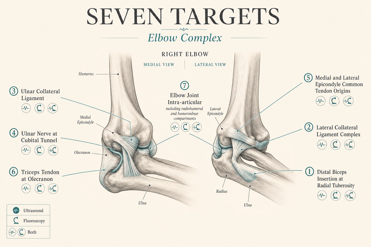
What follows is a section-by-section walk through each structure I target in a comprehensive regenerative elbow procedure. For each one I describe what the structure does when it is healthy, how it manifests as pain when it is not, and how it is targeted — including which imaging tool is right for that specific target.
1. The distal biceps insertion — the tendon at the front of the elbow
What it does: The distal biceps tendon crosses the front of the elbow and inserts on the radial tuberosity, a small bony prominence on the medial side of the proximal radius. Its primary job is not elbow flexion (the brachialis does most of that) but forearm supination — the powerful outward rotation of the palm-up motion. Anyone who has ever turned a stubborn screwdriver or opened a stuck jar has felt the distal biceps working. The tendon inserts through a small bursa (the bicipitoradial bursa) and passes through anatomy that is largely inaccessible to palpation.
How it manifests as pain: Distal biceps tendinopathy or partial tear presents as deep anterior elbow pain, worse with resisted supination and resisted elbow flexion at ninety degrees, and often accompanied by tenderness in the antecubital fossa. In a complete distal biceps rupture the patient hears a pop, develops immediate swelling and bruising, and loses roughly forty to fifty percent of supination strength — that presentation is typically a surgical decision. Partial tears and tendinopathy are the presentations that respond to a well-placed regenerative injection.
How we target it: With an ultrasound-guided peritendinous or intratendinous injection at the distal biceps insertion on the radial tuberosity. This is a target that essentially cannot be done well without ultrasound. The bicipitoradial bursa and the tendon insertion sit deep under muscle bellies with no reliable palpable landmarks (Ultrasound-guided distal biceps anatomy and technique). The evidence for orthobiologic here is small but consistent: in a series of six patients with distal biceps partial tears treated with ultrasound-guided PRP, the mean Mayo Elbow Performance Index rose from 68 to 95, VAS pain with activity fell from 7.25 to 1.3, and all patients returned to full activity (Barker et al., 2015). A second series of twelve patients with an average of forty-seven months of follow-up showed VAS pain with activity falling from 8 to 2.5 with uniform patient satisfaction (Sanli et al., 2016). Ultrasound is the imaging tool for this section without exception.
2. The lateral collateral ligament (LCL) complex — the stabilizer on the outside of the elbow
What it does: The lateral collateral ligament complex is not a single ligament but a fan of three: the radial collateral ligament (RCL), the lateral ulnar collateral ligament (LUCL), and the annular ligament. Together they resist varus and posterolateral rotatory forces on the elbow — the forces that would drive the radial head backward and outward relative to the humerus. The LUCL in particular is the primary restraint against posterolateral rotatory instability, the most common form of chronic elbow instability. When the complex is intact, we do not notice it. When it is disrupted, patients describe the elbow “giving way” on push-ups, on getting out of a chair, or in any position that combines extension with supination.
How it manifests as pain: LCL complex injury presents as lateral elbow pain, a sensation of instability with the arm in extension, positive posterolateral rotatory drawer or table-top relocation tests, and, in more severe cases, frank subluxation events. It is often iatrogenic — the classic mechanism is repeated corticosteroid injection for lateral epicondylitis that weakens the underlying ligament, or a lateral epicondyle release procedure that inadvertently violates the LUCL origin. It is one of the reasons repeated steroid shots for “tennis elbow” can be worse than doing nothing.
How we target it: With ultrasound-guided orthobiologic injection into or along the specific band of the LCL complex. High-frequency ultrasound identifies the RCL, LUCL, and annular ligament as discrete hyperechoic ligamentous bands overlying the radiohumeral joint. PRP or bone marrow concentrate can be placed precisely at the site of ligament thinning or partial disruption. Regenerative treatment is not appropriate for a frankly torn LUCL in a young athlete — that is a surgical reconstruction. But for the far more common presentation of chronic partial LCL laxity in a middle-aged patient with functional instability, targeted ligament injection is a legitimate option and a growing part of orthobiologic practice.
3. The ulnar collateral ligament (UCL) — the pitcher’s ligament
What it does: The ulnar collateral ligament is the medial side stabilizer of the elbow. It resists valgus forces — the outward-bending forces on the elbow — and it is loaded on every overhead throw. In a pitcher, the UCL absorbs peak stresses that come very close to the ligament’s ultimate tensile strength on each pitch. It is the ligament that is reconstructed in “Tommy John” surgery. It is also the ligament that, when partially injured, has become one of the marquee applications of orthobiologic medicine.
How it manifests as pain: UCL injury presents as medial elbow pain in a throwing athlete, loss of velocity, loss of command (particularly location of the fastball), and, on physical exam, tenderness over the ligament and positive valgus stress and moving valgus stress tests. MRI or MR arthrography grades the injury: Type I is a normal-appearing ligament with an inflammatory partial-thickness signal, Type II is a partial-thickness tear, Type III is a complete tear, and Type IV describes a chronically insufficient ligament with attenuation and heterotopic calcification.
How we target it: With ultrasound-guided PRP injection at the site of ligament pathology, sometimes combined with a small amount of orthobiologic into the humeroulnar joint. The evidence in the throwing athlete is now substantial. In an outcome study of ninety-eight professional and elite amateur baseball players stratified by ligament tear grade, orthobiologic injection produced a 61.5 percent return-to-play rate at the prior level for Type I and Type II tears, and a 100 percent return-to-play rate for Type III tears in the appropriate subset — while Type IV tears (already chronically insufficient) returned at only 12.5 percent and should be considered for surgery rather than injection (Chauhan et al., outcomes by MRI grade). A meta-analysis of PRP for partial UCL tears in baseball players demonstrated a pooled return-to-sport rate of approximately seventy-five percent at a mean of eighty-two days — substantially faster than the twelve-to-eighteen-month timeline of a Tommy John reconstruction (Meta-analysis, 2024). Best results occur with leukocyte-poor PRP and a structured twelve-week rehabilitation and throwing program; abbreviated rehabilitation protocols predict reinjury (Technique and complications review, 2023). This is a target where ultrasound is essential — the ulnar nerve sits within millimeters of the ligament, and no fluoroscopic image will keep a needle safely away from the nerve.
4. The ulnar nerve at the cubital tunnel — hydrodissection for cubital tunnel syndrome
What it does: The ulnar nerve is one of the three main nerves of the arm and hand. It travels along the medial side of the arm, wraps around the medial epicondyle, and passes through the cubital tunnel at the elbow on its way to the forearm and hand. It supplies the small muscles of the hand that let us pinch, grip, and spread our fingers, and it carries sensation from the little finger and half of the ring finger. It is second only to the median nerve in frequency of compression neuropathy in the upper limb.
How it manifests as pain: Cubital tunnel syndrome presents as medial elbow pain radiating into the forearm, numbness and tingling in the little and ring fingers, weakness of grip and pinch strength, and, in more severe cases, atrophy of the small muscles of the hand. Symptoms are often positional — worse with the elbow held in flexion, worse at night when sleeping with the elbow bent, worse with prolonged phone use or typing. It is one of the most common reasons a patient with medial elbow pain has had every ligament imaged and every tendon injected without relief — because the actual pain generator is the nerve, not the ligament.
How we target it: With ultrasound-guided hydrodissection of the ulnar nerve at the cubital tunnel. Hydrodissection means using a fluid (typically five percent dextrose in water, or platelet lysate) to gently separate the nerve from the surrounding fascia and scar tissue that has developed around it. It restores the natural gliding motion of the nerve and, in the process, decompresses the nerve without surgery. A three-patient case series using platelet lysate combined with D5W for cubital tunnel syndrome documented 75 percent or greater improvement in pain and function scores and greater than 80 percent single-assessment numeric evaluation scores at follow-up (Platelet lysate hydrodissection series, 2024). Case reports of D5W hydrodissection for cubital tunnel syndrome have documented complete symptom resolution accompanied by normalization of nerve architecture on follow-up MRI (Stoddard 2019, CSMR). Systematic reviews of nerve hydrodissection for entrapment neuropathies have found the technique to be safe and effective across multiple compression sites (Hydrodissection systematic review). In throwing athletes with UCL injury, cubital tunnel symptoms often coexist with medial elbow pain and can be treated in the same session as UCL orthobiologic therapy — a genuine two-structure procedure. This is another target where ultrasound is essential; the ulnar nerve is invisible to fluoroscopy.
5. The medial and lateral epicondyle common tendons — golfer’s and tennis elbow
What they do: The medial epicondyle is the origin of the common flexor tendon — the shared tendinous origin of the muscles that flex the wrist and pronate the forearm. The lateral epicondyle is the origin of the common extensor tendon — the shared origin of the wrist and finger extensors, with the extensor carpi radialis brevis being the tendon most commonly implicated in lateral epicondylitis. These are the two most-injured tendons in the human elbow. Every gripping motion of the hand loads them.
How they manifest as pain: Lateral epicondylitis (“tennis elbow”) presents as lateral elbow pain, tenderness over the lateral epicondyle, and pain with resisted wrist extension and gripping. Medial epicondylitis (“golfer’s elbow”) presents as medial elbow pain, tenderness over the medial epicondyle, and pain with resisted wrist flexion and pronation. Both are technically tendinoses rather than tendinitises — the pathology is collagen disorganization and neovascularization rather than acute inflammation, which is why anti-inflammatory treatments alone often fail and why corticosteroid injections may relieve pain for a few weeks but predict worse two-year outcomes than doing nothing.
How we target them: With ultrasound-guided PRP injection into the site of tendinopathy at the common tendon origin. This is one of the most-studied applications of orthobiologic medicine in the elbow. A 2025 randomized controlled trial published in the American Journal of Sports Medicine followed patients with lateral epicondylitis treated with PRP, extracorporeal shockwave therapy, or physical therapy alone for two years; DASH score improvement was 31 points with PRP versus 19 points with physical therapy and 18 points with shockwave, with patient satisfaction of 4.6 out of 5 for PRP versus 3.0 for physical therapy and 3.2 for shockwave — a durable two-year superiority (Lhee et al., AJSM 2025). A 2024 randomized trial comparing PRP with dry needling in lateral epicondylitis found PRP superior on all outcome measures at nine months (PRP vs. dry needling RCT). More recent randomized work has found that PRP, corticosteroid, and ozone injections all produce clinically meaningful improvement in lateral epicondylitis with no clear superiority among them at short-term follow-up — but the durability of PRP appears greater on longer follow-up (Multicenter RCT, 2025). Precision matters. Blind injections at the medial epicondyle risk the ulnar nerve; blind injections at the lateral epicondyle can miss the pathologic tissue that is often deeper than expected. Ultrasound is essential.
6. The triceps tendon at the olecranon — the posterior elbow’s workhorse
What it does: The triceps is the powerful three-headed extensor of the elbow. Its shared tendon inserts on the olecranon, the bony prominence at the back of the elbow. The triceps straightens the elbow against resistance — every push-up, every bench press, every push of a heavy door depends on it. Compared to the common flexor and extensor tendons at the epicondyles, the triceps tendon is a stronger, thicker structure, but it is not immune to tendinopathy or partial tear.
How it manifests as pain: Triceps tendinopathy presents as posterior elbow pain, tenderness over the olecranon insertion, and pain with resisted elbow extension. In weightlifters and in patients with certain systemic risk factors it can progress to partial or complete tendon tear — the classic mechanism is a heavy bench press with the elbow extended against maximal resistance. Complete tears require surgical repair. Partial tears and tendinopathy respond to orthobiologic therapy.
How we target it: With ultrasound-guided PRP or bone marrow concentrate injection at the site of triceps tendinopathy or partial tear. Ultrasound identifies the triceps tendon as it inserts on the olecranon and clearly shows the location and depth of any fiber disruption. The biologic is placed precisely at the pathology, not blindly into the general area. This is soft tissue at moderate depth against a bony insertion — ultrasound is the right tool.
7. The elbow joint compartments — humeroulnar and radiohumeral intra-articular injections
What they do: The elbow contains three articulations within a single capsule: the humeroulnar joint (the hinge between the humeral trochlea and the ulnar trochlear notch), the radiohumeral joint (the ball-and-socket-like articulation between the capitellum and the radial head), and the proximal radioulnar joint (the pivot that allows forearm pronation and supination). Cartilage lines all three, and all three communicate as a single joint space. Osteoarthritis, inflammatory arthritis, and post-traumatic arthritis can affect any of these articulations, most commonly the radiohumeral joint after radial head fracture or the humeroulnar joint after major elbow trauma.
How they manifest as pain: Elbow joint pain presents as diffuse deep elbow pain, worse with rotation and end-range motion, worse with weight-bearing (leaning on the arm), and often accompanied by stiffness, crepitus, and loss of terminal extension. In advanced disease, patients cannot fully extend or flex the elbow, and simple tasks like carrying a briefcase become uncomfortable. This is the joint most often targeted for intra-articular corticosteroid, hyaluronic acid, or biologic therapy.
How we target it: With an intra-articular elbow injection under ultrasound, with fluoroscopic arthrography added when confirmation of intra-articular flow or a specific compartment target is required. Ultrasound-guided posterior or lateral approaches into the elbow joint are highly accurate in trained hands and are the standard for most patients. When the diagnosis benefits from arthrographic confirmation — for example, when a specific compartment is being targeted, when we want to confirm that a communicating tear allows fluid to flow between compartments, or when the joint is severely degenerated with tight capsular constraint — we add fluoroscopy with a small amount of iodinated contrast to visualize joint flow. This is one of the few elbow targets where both imaging tools are used together, and using both is what turns a good injection into a diagnostic and therapeutic one.
How the conductor puts the elbow ensemble together
A comprehensive regenerative elbow procedure at Pravida does not treat all seven structures on every patient. The point of the physical exam, the imaging review, and the diagnostic history is to determine which sections of the ensemble are actually out of tune for you. Some patients have a single-structure problem — an isolated lateral epicondylitis, an isolated distal biceps tendinopathy — and a single well-placed injection is exactly the right answer. Other patients have two or three structures contributing simultaneously (the most common combined pattern in throwers is UCL plus ulnar nerve; in middle-aged patients with a history of repeated cortisone injections it is common flexor or extensor tendinopathy plus underlying ligament laxity), and treating just one of them is precisely why the last three injections wore off in weeks.
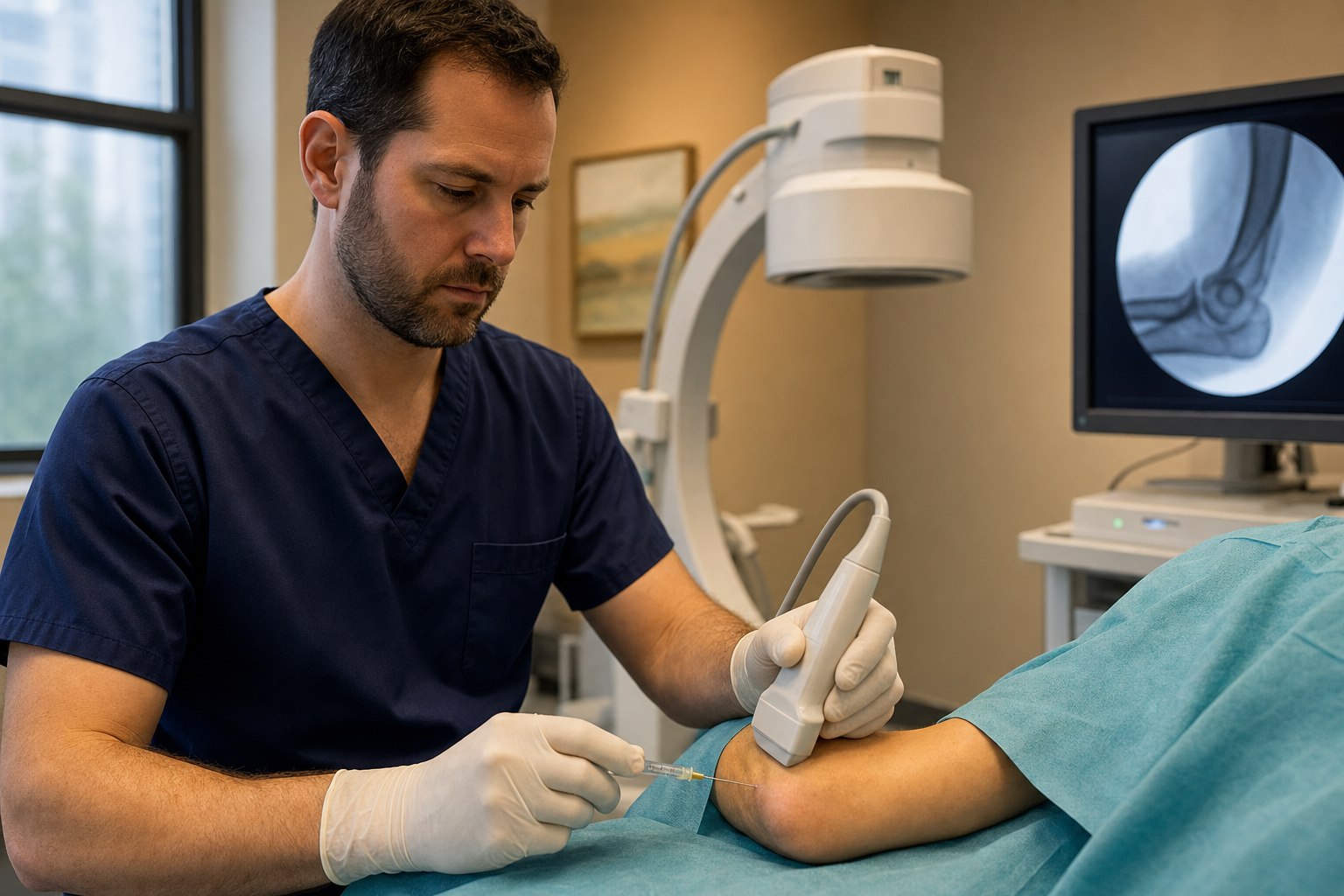
What a world-class elbow procedure looks like on the day it happens: a physical exam and imaging review that identifies the specific structures in play, a written plan for which structures will be treated, a procedure suite set up with both a C-arm fluoroscope and a high-resolution ultrasound machine, and a sequence of small, precise injections — each one confirmed on real-time imaging before medication is delivered — that treats each identified section of the ensemble with the tool that shows it best. On the medial side, ultrasound is the exclusive imaging tool. On the joint itself, fluoroscopy may be added for confirmation. Most elbow procedures are outpatient and take under an hour. Most patients drive themselves home.
The choice of biologic — platelet-rich plasma (typically leukocyte-poor for tendon and ligament work), bone marrow concentrate for higher-grade partial tears, or in some cases a targeted non-regenerative agent for a specific structure — is guided by the tissue being treated and by the patient’s overall regenerative plan. The CartiNova program is our organized framework for making those choices in a way that is transparent, evidence-informed, and personalized to the individual patient.
What most elbow patients get wrong before they see us
- They accept “tennis elbow” as the whole diagnosis. Lateral epicondylitis is a real thing, but so is an underlying LCL laxity that repeated cortisone shots have quietly created. When “the tennis elbow shot” is no longer working, the answer is usually not another shot — it is a broader diagnostic look that includes the ligament, the radiohumeral joint, and the specific tendon fibers involved.
- They forget the nerve on the medial side. Medial elbow pain is not always the UCL and not always the common flexor tendon. In a substantial fraction of patients — particularly those with paresthesias, night symptoms, or a history of repetitive elbow flexion — the ulnar nerve is the actual pain generator. Ignoring it is why some medial elbow “golfer’s elbow” injections never work.
- They accept fluoroscopic injections on the medial side. Any medial elbow soft tissue procedure — UCL, common flexor tendon, ulnar nerve — needs ultrasound to keep the needle away from the ulnar nerve. An injection performed without direct nerve visualization is an unnecessary risk.
- They accept repeated cortisone injections as maintenance therapy. Corticosteroid can produce meaningful short-term relief in lateral or medial epicondylitis, but repeated injections weaken the underlying tendon and ligament and predict worse two-year outcomes than watchful waiting. Orthobiologic therapy has the opposite trajectory: modest early benefit, durable and sometimes growing benefit over time.
- They expect regeneration without rehabilitation. A biologic injection into a compromised tendon does not rebuild strength on its own. It creates a window in which targeted eccentric loading and progressive strengthening can rebuild the tendon. In the thrower, best results consistently follow a structured twelve-week rehabilitation and interval throwing program. The injection and the rehab are one intervention, not two.
The honest limits, and where a good clinician still matters
- The seven-structure approach is designed for patients with chronic or subacute mechanical elbow pain that has not fully resolved with conservative care. It is not first-line treatment for acute traumatic complete ruptures in surgical candidates (complete distal biceps tears, high-grade UCL tears in a young elite thrower who has already decided on reconstruction, complete triceps ruptures), for elbow instability requiring reconstruction, or for red-flag presentations.
- Type IV UCL injury — a chronically insufficient, attenuated, and calcified ligament — is a poor candidate for orthobiologic therapy and should typically be considered for surgical reconstruction. Patient selection matters, and not every partial UCL injury is treatable by injection.
- Any medial elbow procedure requires ultrasound visualization of the ulnar nerve. If a clinician offers a fluoroscopically-guided-only injection at the medial elbow, that is a workflow issue, not a matter of preference.
- Regenerative outcomes depend on the underlying tissue, the biologic used, the technical precision of delivery, and the rehabilitation that follows. A biologic delivered blindly, or into the wrong structure, is not regenerative — it is just an injection.
- If you have new hand weakness with visible muscle wasting, sudden loss of forearm rotation after trauma, a joint that will not stay in place, unexplained fever, or elbow pain accompanied by symptoms of a cardiac or vascular problem, please contact your physician or an emergency department today. Those are not situations for an elective regenerative procedure.
Ready to have your specific elbow pattern mapped?
If you are an Atlanta-area patient with chronic elbow pain that has not fully responded to prior injections or conservative care — or a thrower who wants a candid, evidence-informed second opinion on whether orthobiologic therapy is appropriate for your UCL injury — we would rather see you in person. A consultation at Pravida Health includes a physical exam, an imaging review, and, where appropriate, a comprehensive regenerative plan tailored to the specific sections of your elbow ensemble that are out of tune.
Book a consultationKey sources referenced in this article
- Ultrasound anatomy and technique for distal biceps injection. PMC8181841. Details the sonographic identification of the bicipitoradial bursa and distal biceps insertion at the radial tuberosity, structures that lack reliable palpable landmarks.
- Barker SL, et al. Ultrasound-guided PRP for distal biceps partial tears. PMC4935115. Case series of six patients with Mayo Elbow Performance Index rising from 68 to 95 and VAS activity pain from 7.25 to 1.3.
- Sanli I, et al. Long-term follow-up of PRP for distal biceps tendinopathy. PMC4935121. Twelve patients with mean 47-month follow-up; VAS activity from 8 to 2.5 with uniform patient satisfaction.
- Chauhan A, et al. Outcomes of PRP for UCL injury in baseball players by MRI tear grade. PMC7983438. Type I/II 61.5% return to play, Type III 100%, Type IV 12.5% (surgical candidate).
- Meta-analysis: PRP for partial UCL tears in baseball players. PMC11418690. Pooled return-to-sport rate of approximately 75 percent at a mean of 82 days.
- UCL PRP technique review and complications. PMC10426667. Leukocyte-poor PRP with a structured 12-week rehabilitation and throwing program; ulnar nerve fibrosis reported with fluoroscopic technique and subluxing ulnar nerves.
- Case report and technique review: ulnar nerve injury with fluoroscopic-only medial elbow injection and rescue with ultrasound-guided hydrodissection. PMC10426644. Illustrates the safety case for mandatory ultrasound on the medial side.
- Stoddard 2019: D5W hydrodissection for cubital tunnel syndrome. Current Sports Medicine Reports. Full symptom resolution with normalization of nerve architecture on follow-up MRI.
- Platelet lysate plus D5W hydrodissection for cubital tunnel syndrome. PMC11954682. Three-patient series with 75 percent-plus pain and function improvement and greater than 80 percent SANE scores.
- Lhee et al., AJSM 2025: PRP vs. shockwave vs. physical therapy for lateral epicondylitis at two years. Summary in clinical practice review. Clinical summary of AJSM 2025 RCT. DASH improvement 31 (PRP) vs. 19 (PT) vs. 18 (shockwave); satisfaction 4.6 vs. 3.0 vs. 3.2.
- Randomized controlled trial: PRP vs. dry needling for lateral epicondylitis at nine months. PMC11178710. PRP superior on pain, function, and grip strength outcomes.
- Multicenter randomized trial: PRP, corticosteroid, and ozone for lateral epicondylitis. PMC13078189. All three produce clinically meaningful improvement at short-term follow-up.