
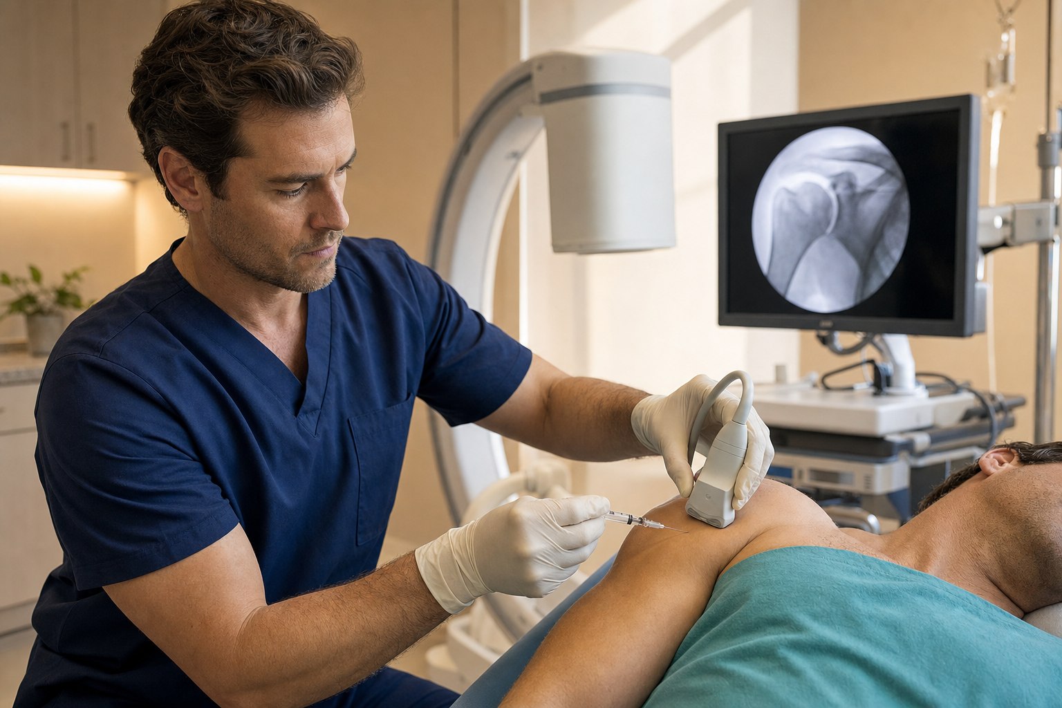
The shoulder as an orchestra, not a hinge
The shoulder is the most mobile joint in the human body, and every ounce of that mobility is bought at the cost of stability. Unlike the hip, which sits deep inside a bony socket, the humeral head sits on the glenoid the way a golf ball sits on a tee — almost entirely dependent on soft tissue to stay put and move smoothly. That soft tissue is not one thing. It is an orchestra of tendons, ligaments, bursae, cartilage, labrum, capsule, and the peripheral nerves that report on all of it in real time. When one section falls out of tune, the whole ensemble sounds off — and the pain does not always come from where the injury lives.
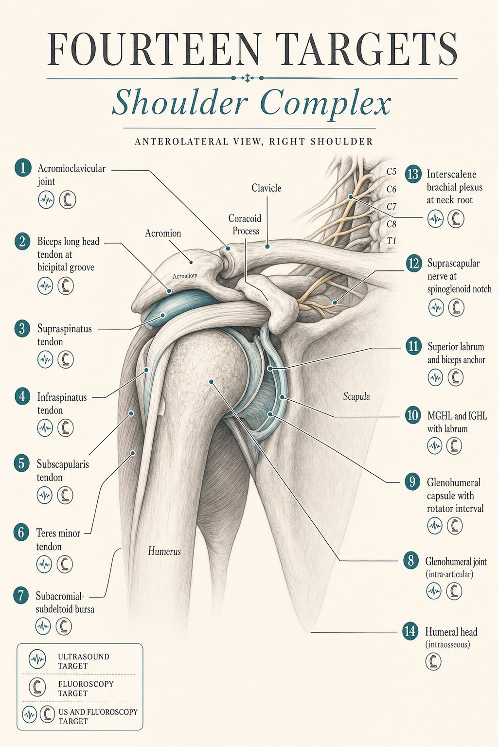
This is the second article in a series on how a master regenerative procedure actually works, region by region. In the first article we walked through the five-structure ensemble of the lumbar spine. The shoulder is a more complex instrument. In my hands, a comprehensive regenerative shoulder procedure has as many as fourteen distinct anatomic targets, each with its own indication, its own imaging modality, and its own role in the ensemble. Most shoulder patients do not need all fourteen. But almost every shoulder patient who has “had a shot that stopped working” has had one or two structures treated when three or four were the actual problem.
What follows is the conductor’s map: every structure I treat in the shoulder, why it matters, how it produces pain when it is out of tune, and which imaging tool — ultrasound, fluoroscopy, or both — is right for that particular section.
Why two imaging tools, and not just one
The same principle we applied to the lumbar spine applies here, only with different structures. Ultrasound is real-time sound imaging. It cannot see through bone, but it excels at showing tendon fibers, bursal fluid, ligament architecture, capsular thickness, labral edges at the periphery, peripheral nerves, and, most importantly, a needle moving through all of them in real time. For soft tissue and peripheral nerve work in the shoulder, ultrasound is the standard of care.
Fluoroscopy is real-time X-ray. It shows bone, and with a small amount of contrast dye it shows the flow of medication through spaces that live deep against bone — the true intra-articular space of the glenohumeral joint on arthrography, the interior of the humeral head for subchondral or intraosseous work, and the pattern of capsular filling during hydrodilatation. Every well-trained interventionalist can inject a glenohumeral joint under ultrasound alone. But when the diagnosis is arthrographic (Is this capsule contracted? Is that dye going where I think it is?) or when the target lives inside the bone, fluoroscopy is what confirms it.
Neither tool is a substitute for the other. A physician who uses only ultrasound has to work blind on any intraosseous target and cannot arthrographically confirm capsular volume. A physician who uses only fluoroscopy cannot see the difference between a partial supraspinatus tear and a full-thickness one at the point of the needle. A physician who uses both can pick the right tool for each of the fourteen sections of the ensemble.
The fourteen sections of the shoulder orchestra
What follows is a section-by-section walk through each structure I target in a comprehensive regenerative shoulder procedure. For each one I describe what the structure does when it is healthy, how it manifests as pain when it is not, and how it is targeted — including which imaging tool is right for that specific target.
1. The acromioclavicular (AC) joint — the small joint at the top of the shoulder
What it does: The AC joint is the small articulation where the outer end of the clavicle meets the acromion of the scapula. It is a low-motion, gliding joint that acts as a suspension linkage — it allows the scapula to rotate freely on the collarbone during overhead motion while transmitting the load of the arm back through the clavicle to the trunk. It is a tiny joint that carries a huge amount of leverage.
How it manifests as pain: AC joint pain is characteristically pinpoint. Patients can put a fingertip directly on the joint and reproduce their pain. It is worse with cross-body adduction (reaching across the chest for a seatbelt), overhead pressing, and lying on the affected side. Because it sits so close to the subacromial bursa and the supraspinatus tendon, AC joint pain is one of the most misidentified causes of “impingement” symptoms — a patient with a symptomatic AC joint may have every impingement test positive and every rotator cuff test unremarkable, and the diagnosis is confirmed only when a small volume of anesthetic into the AC joint completely abolishes the pain.
How we target it: With an intra-articular AC joint injection under ultrasound and, when needed, fluoroscopic confirmation. The joint is small (often only a few millimeters wide) and its plane can be angled fifteen to twenty degrees off vertical. Ultrasound identifies the joint capsule and confirms intra-articular needle position by watching the fluid distend the joint. Fluoroscopy with a drop of contrast is used when the joint is severely degenerated or when a diagnostic block is being placed to definitively separate AC joint pain from rotator cuff pain. Both tools together give a level of certainty that neither alone provides.
2. The biceps long head tendon — the intra-articular tendon at the front of the shoulder
What it does: The long head of the biceps is unusual: it is a tendon that runs inside the glenohumeral joint before it exits through a narrow bony groove (the bicipital groove) on the front of the humerus. Its role in shoulder mechanics is debated and probably modest — but its role as a pain generator is not modest at all. The tendon is heavily innervated, sits in a bony tunnel under a retinacular sling, and is one of the most consistent sources of anterior shoulder pain that patients localize with a fingertip to the front of the humeral head.
How it manifests as pain: Biceps tendinopathy or partial tear presents as anterior shoulder pain worse with resisted elbow flexion, resisted forearm supination (Yergason’s test), overhead reaching, and lying on the affected side. On palpation the bicipital groove is tender. In more advanced disease the tendon can become frankly torn or can dislocate medially out of the groove, producing a mechanical popping sensation. Biceps pathology travels with rotator cuff pathology far more often than either travels alone — which is one of the reasons treating only one of the two can leave a shoulder incompletely treated.
How we target it: With ultrasound-guided injection into the biceps tendon sheath for tendinopathy or partial tear, and with peritendinous orthobiologic injection where the tendon shows disruption or hypoechoic degeneration. Ultrasound is uniquely powerful here: it shows the tendon in cross-section within the bicipital groove, it shows tendon subluxation dynamically with rotation, and it lets us place a needle around (never into) the tendon substance to bathe it in a biologic without penetrating an already compromised fiber structure. This is a target that essentially cannot be done well without ultrasound.
3. The supraspinatus tendon — the roof of the rotator cuff
What it does: The supraspinatus is the top-most muscle of the rotator cuff. Its tendon crosses the top of the humeral head and inserts on the greater tuberosity. It initiates abduction of the arm and, throughout arm elevation, holds the humeral head centered on the glenoid so that the deltoid can lift the arm without the head migrating upward. It is the single most-injured tendon in the human shoulder, largely because of where it sits: in a tight subacromial space, exposed to compression, and with a well-documented critical zone of decreased blood supply.
How it manifests as pain: Supraspinatus pathology is the classic “painful arc” — discomfort with active abduction between sixty and one hundred twenty degrees, weakness with resisted abduction in scaption (the empty-can test), and lateral shoulder pain that patients often localize with a hand cupped over the deltoid rather than a single fingertip. Sleep on the affected side is often the first casualty. Partial-thickness tears are common in patients over forty and are frequently the pain generator behind a shoulder that has “been bothering me for a year.”
How we target it: With ultrasound-guided orthobiologic injection to the site of partial tear or tendinopathy, typically platelet-rich plasma placed directly at the footprint or into the disrupted fibers. The evidence here is meaningful. In a randomized trial of partial-thickness supraspinatus tears, PRP produced significantly greater tear size reduction at six months than corticosteroid (an average of 3.39 mm versus 1.10 mm), along with better pain and function scores (Randomized trial, 2023). Longer-term work has shown that ultrasound-guided PRP into the rotator cuff delays the progression of degenerative changes compared to medical therapy alone (Arrigoni et al., RSNA). For calcific tendinopathy specifically, ultrasound-guided barbotage of the calcification followed by PRP has shown complete healing rates approaching eighty percent at six months (Calcific tendinopathy series). Ultrasound is the imaging tool for this section without exception.
4. The infraspinatus tendon — the external rotator behind the shoulder
What it does: The infraspinatus originates from the back surface of the scapula and inserts on the greater tuberosity just behind the supraspinatus. Its job is external rotation of the arm and dynamic posterior stabilization of the humeral head against the glenoid. Every time you brush your hair, throw a ball, or reach behind your back, the infraspinatus is one of the muscles that lets you do it.
How it manifests as pain: Infraspinatus tendinopathy or partial tear presents as posterior shoulder pain, weakness with resisted external rotation (the arm at the side, the elbow at ninety degrees, and a fight against a hand pushing inward), and pain with reaching behind the back. It is under-diagnosed compared to supraspinatus pathology, largely because it is behind rather than on top of the shoulder and does not produce a classic painful arc. But in throwing athletes and in patients with a chronic posterior shoulder ache, the infraspinatus is often the actual pain generator.
How we target it: With ultrasound-guided orthobiologic injection into the tendon substance or at the tendon footprint. Ultrasound clearly identifies the infraspinatus fibers as they course laterally to insert on the greater tuberosity, and the same principles that apply to the supraspinatus apply here — PRP or bone marrow concentrate placed precisely at the site of pathology, under real-time needle visualization. This is soft tissue at moderate depth. Ultrasound is the appropriate tool.
5. The teres minor tendon — the small external rotator often forgotten
What it does: The teres minor is the small muscle immediately below the infraspinatus, sharing the job of external rotation and posterior stabilization. It has its own tendon insertion on the greater tuberosity and its own nerve supply (the axillary nerve). It is small, it is often overlooked, but in specific patient populations — particularly patients with quadrilateral space syndrome or with isolated axillary neuropathy — it is the tendon that fails first.
How it manifests as pain: Isolated teres minor pathology is uncommon but not rare. It presents as posterior shoulder pain and specific weakness with the elbow tucked to the side and the arm in external rotation at ninety degrees of shoulder elevation — a position that isolates teres minor from infraspinatus. On MRI the teres minor may show selective fatty infiltration when its axillary nerve supply has been compromised. When conventional shoulder injections have not worked and posterior shoulder pain persists, the teres minor is one of the structures the ensemble may be missing.
How we target it: With ultrasound-guided orthobiologic injection at the teres minor footprint or into its tendon substance. The teres minor is exquisitely well-visualized on ultrasound, sitting immediately inferior to the infraspinatus with its characteristic short, fan-shaped insertion. This is another target where ultrasound is essentially the only option.
6. The subscapularis tendon — the internal rotator at the front
What it does: The subscapularis is the fourth muscle of the rotator cuff and the only one on the front of the scapula. Its broad tendon crosses the front of the humeral head and inserts on the lesser tuberosity. It is the primary internal rotator of the arm and the anterior counterbalance to the posterior cuff. Everything from reaching into your back pocket to lifting a heavy object off a table depends on it firing correctly.
How it manifests as pain: Subscapularis pathology is under-recognized. Classical teaching has focused on the posterior-superior cuff (supraspinatus, infraspinatus) for decades, but the subscapularis is the pain generator in more shoulders than most patients (and many physicians) realize. It presents as anterior shoulder pain, weakness with the lift-off test (the hand behind the back, lifted away from the low back against resistance), and, in more advanced pathology, medial subluxation of the biceps tendon as its retinacular sling from the subscapularis fails. When a patient has anterior shoulder pain that has been called “biceps tendinitis” for years without responding to biceps-focused treatment, the subscapularis is often the reason.
How we target it: With ultrasound-guided orthobiologic injection to the subscapularis tendon or footprint. Ultrasound is the only imaging tool that shows the subscapularis tendon in cross-section as the arm is passively externally rotated — a maneuver that unfurls the tendon and lets us examine every fiber in a way even MRI cannot dynamically match. Precision here is important because the tendon lives just millimeters from the coracoid process and the intra-articular space.
7. The subacromial-subdeltoid bursa — the lubricating layer under the roof
What it does: The subacromial-subdeltoid bursa is a thin lubricating sac that sits between the deep surface of the deltoid and acromion above and the rotator cuff tendons below. It allows the cuff to glide smoothly under the acromion as the arm elevates. When it is healthy, it is a nearly invisible film of synovial fluid. When it is inflamed, it can be one of the loudest sections in the shoulder orchestra.
How it manifests as pain: Subacromial bursitis presents as diffuse lateral shoulder pain, worse with any overhead reaching, worse at night when lying on the affected side, and often accompanied by a full painful arc. It is one of the most common initial diagnoses in patients with shoulder pain and one of the most common structures targeted by a first-line shoulder injection. In an acute inflammatory bursitis a single well-placed injection can produce dramatic relief. The problem is when the bursitis is chronic, secondary to an underlying rotator cuff or AC joint pathology — a repeat bursa injection every few months without treating what is actually driving the bursal inflammation is one of the most common patterns of an incompletely treated shoulder.
How we target it: With ultrasound-guided injection directly into the bursa. Ultrasound identifies the bursa as a hypoechoic band between the deltoid and the supraspinatus tendon and confirms intrabursal needle position by watching the injectate spread within the bursal plane. Blind subacromial injections have been shown to be intrabursal only about sixty to seventy percent of the time; ultrasound-guided injections approach one hundred percent. When the bursitis is secondary to an underlying cuff problem, we treat the bursa and the cuff in the same session — that is the ensemble at work.
8. The glenohumeral joint (intra-articular) — the main ball-and-socket
What it does: The glenohumeral joint is the true ball-and-socket of the shoulder — the articulation between the humeral head and the glenoid of the scapula, lined with articular cartilage and enclosed by the joint capsule. It is the joint responsible for the vast majority of shoulder motion. Its cartilage can wear (glenohumeral osteoarthritis), and its intra-articular space communicates with the biceps sheath and, in adhesive capsulitis, with a pathologically contracted capsule.
How it manifests as pain: Glenohumeral joint pain presents as a deep, diffuse ache in the shoulder, worse with rotation in either direction, worse with weight-bearing (leaning on the arm), and often accompanied by stiffness and crepitus. In advanced osteoarthritis the range of motion is globally reduced — both active and passive — and simple tasks like reaching to a high shelf become impossible. This is the joint most often targeted for an intra-articular corticosteroid or biologic injection.
How we target it: With an intra-articular glenohumeral injection under ultrasound, with fluoroscopic arthrography added when the diagnosis or the space requires it. Ultrasound-guided posterior approach into the glenohumeral joint is highly accurate in trained hands. For patients where the capsule may be contracted or where we need to document intra-articular flow of a specific volume (as in hydrodilatation, described below), we add fluoroscopy with a small amount of iodinated contrast and observe the classic arthrographic pattern of the joint. This is one of the few targets where both imaging tools are used in a single procedure, and using both is what turns a good injection into a diagnostic and therapeutic one.
9. The glenohumeral capsule and the rotator interval — hydrodilatation for adhesive capsulitis
What it does: The glenohumeral joint capsule is a fibrous sleeve that surrounds the joint and allows its enormous range of motion by being long, elastic, and freely mobile. The rotator interval is a small triangular window in the front of the capsule between the supraspinatus above and the subscapularis below, containing the coracohumeral ligament, the superior glenohumeral ligament, and the biceps tendon. When the capsule works, it goes almost unnoticed. When it fails, it becomes the loudest section in the shoulder.
How it manifests as pain: Adhesive capsulitis — the true “frozen shoulder” — is a distinctive pattern of capsular thickening and contraction that produces global loss of active and passive range of motion, with external rotation typically the most severely restricted. Patients cannot lift the arm past shoulder height and cannot allow another person to lift it for them either. The pain phase is often intense, especially at night, and gradually gives way to a stiff phase that can last months. The underlying pathology is a genuine contracture of the capsule, not simply a weak or inhibited muscle.
How we target it: With ultrasound-guided (and, where indicated, fluoroscopically confirmed) capsular hydrodilatation through the rotator interval. Hydrodilatation is not just an intra-articular injection — it is the deliberate stretching of a contracted capsule from the inside, by injecting a larger volume of solution (typically twenty to forty milliliters) than the pathologically small joint can hold, until the capsule is dilated and, in some cases, ruptured in a controlled manner. Meta-analysis of randomized trials shows that hydrodistension significantly improves pain and function compared to conventional treatment in adhesive capsulitis (Meta-analysis, Medicine 2024). An anterior approach through the rotator interval has been shown to be superior to a posterior approach for pain relief and range of motion (Comparative study, 2020), and ultrasound guidance outperforms fluoroscopy alone for both accuracy and outcomes (Ultrasound vs. fluoroscopy comparison). In practice, we most often combine hydrodilatation with a suprascapular nerve block and, when appropriate, a supervised rehabilitation program (Combined therapy series) — the injection is one element of a broader plan, never a standalone treatment.
10. The MGHL, IGHL, and posterior capsule with the labrum — the ligamentous restraints
What they do: The glenohumeral joint has three named intracapsular ligaments — the superior, middle, and inferior glenohumeral ligaments (SGHL, MGHL, IGHL) — each of which becomes taut in a specific arm position to prevent excessive translation of the humeral head. The SGHL tightens with the arm in adduction. The MGHL tightens at approximately forty-five degrees of abduction with external rotation. The IGHL complex is the strongest static stabilizer and becomes the primary restraint against anterior-inferior translation at ninety degrees of abduction with external rotation — the classic apprehension position (Ligament biomechanics review, University of Washington orthopedics anatomy reference). The labrum is a fibrocartilaginous rim around the glenoid that deepens the socket and provides the attachment point for these ligaments. The posterior capsule, when tight, produces a specific pattern of loss of internal rotation known clinically as GIRD (glenohumeral internal rotation deficit).
How they manifest as pain: Ligamentous laxity or partial injury of the anterior band of the IGHL presents as anterior shoulder apprehension, a sensation of the shoulder wanting to slip forward in specific positions, and pain with the arm in the ninety-ninety abducted, externally rotated position. Posterior capsule tightness presents as loss of internal rotation, posterior shoulder pain in throwers, and secondary rotator cuff symptoms as the humeral head migrates posterosuperiorly with each throw. These are common patterns in overhead athletes, in patients with subtle multidirectional instability, and in patients with a labral tear that has destabilized the associated ligament.
How we target them: With ultrasound-guided injection into the specific ligament or the ligament-labrum junction. High-frequency ultrasound identifies the anterior and inferior joint capsule with its ligamentous thickening, and orthobiologic can be placed precisely along the ligament to support healing of a partial ligament injury or the periligamentous capsular tissue. For posterior capsule tightness, ultrasound-guided hydrodissection between the posterior capsule and the posterior deltoid can release restricted glide. This is soft-tissue work at the periphery of the joint — ultrasound is essential.
11. The superior labral anchor and biceps origin — the SLAP zone
What it does: The superior labrum is the top portion of the glenoid labrum, and it is the anchor point where the long head of the biceps tendon originates. Together, the superior labrum and biceps anchor act as one biomechanical unit that resists distraction and rotation of the humeral head, especially in overhead athletes. When the anchor is intact, the biceps and the superior glenoid work as one.
How it manifests as pain: A SLAP (Superior Labrum Anterior to Posterior) tear is a distinctive injury pattern in throwers, patients with a fall on an outstretched arm, and older adults with a chronic degenerative version. It presents as deep, poorly-localized shoulder pain, mechanical catching, pain with the arm in overhead throwing positions, and often positive active compression and biceps load tests. Treatment decisions in SLAP tears are age-stratified: primary labral repair is favored in patients under forty, and biceps tenodesis (releasing the biceps anchor and reattaching the tendon lower on the humerus) is favored in patients over forty (SLAP treatment review). Between the surgical options, well-selected nonoperative regenerative treatment has a role in specific patients.
How we target it: With ultrasound-guided orthobiologic injection to the superior labrum and biceps anchor, with fluoroscopic arthrography to confirm intra-articular flow when the diagnosis or position is uncertain. This is a target where both imaging tools shine: ultrasound sees the labral edge and the biceps tendon at their peripheral origin, and fluoroscopy with contrast confirms intra-articular versus extra-articular position when the anchor is being targeted directly. The role of orthobiologic here is not to substitute for surgery in a patient who needs it — it is to give a targeted regenerative option in patients whose imaging findings and clinical picture make them appropriate candidates for a conservative regenerative trial before any surgical decision is made.
12. The suprascapular nerve at the spinoglenoid notch — the peripheral nerve block for shoulder pain
What it does: The suprascapular nerve is a mixed motor-sensory nerve that arises from the upper trunk of the brachial plexus, travels laterally across the top of the scapula, and passes through the suprascapular notch and around the spinoglenoid notch to supply the supraspinatus, the infraspinatus, and, critically, most of the sensory innervation of the glenohumeral joint and the posterior shoulder capsule. About seventy percent of shoulder sensory input travels through this one nerve.
How it manifests as pain: The suprascapular nerve is not a source of pain per se in most patients — it is the route pain travels. Which is exactly why blocking it is so useful. A well-placed suprascapular nerve block can dramatically reduce pain from glenohumeral osteoarthritis, adhesive capsulitis, chronic rotator cuff pathology, and post-surgical shoulder pain. Meta-analyses of randomized trials in chronic shoulder pain show that suprascapular nerve blockade produces standardized mean differences in pain scores in excess of two — a very large clinical effect — with sustained benefit over weeks to months (Chronic shoulder pain meta-analysis).
How we target it: With ultrasound-guided suprascapular nerve block at the spinoglenoid notch (or, less commonly, at the classic suprascapular notch). Ultrasound identifies the nerve as a small hyperechoic structure in a fat-lined groove at the top of the scapula, and a small volume of local anesthetic (with or without a longer-acting agent) is deposited precisely along the nerve. Unlike an interscalene block, the suprascapular block preserves lung function and does not paralyze the diaphragm, which is why an anterior suprascapular approach is increasingly preferred in patients where phrenic sparing matters (Anterior SSB phrenic sparing study). In our practice, the suprascapular block is often used in the same procedure as an intra-articular injection or a hydrodilatation to give the patient meaningful post-procedure pain relief while the biologic gets to work.
13. The interscalene brachial plexus — regional anesthesia for the whole shoulder
What it does: The brachial plexus is the network of nerves that arises from the cervical spine, crosses under the collarbone, and supplies the entire upper limb. At the level of the neck root, between the anterior and middle scalene muscles, the plexus is composed of roots and trunks that can be blocked as a single target with a small volume of local anesthetic. This is the interscalene block — the gold-standard regional anesthetic for shoulder surgery and, in select cases, for extended-duration analgesia in complex regenerative procedures.
How it manifests as pain: Like the suprascapular nerve, the brachial plexus is a route rather than a source. But in patients undergoing extensive regenerative work — multi-structure procedures, hydrodilatation with mobilization, patients with severe procedure-related anxiety — the ability to place a temporary block that completely anesthetizes the shoulder for six to eighteen hours changes the entire experience. The block also serves as a diagnostic tool: if a shoulder pain does not resolve with a well-placed interscalene block, the source is not in the arm or shoulder territory. That is diagnostic information.
How we target it: With ultrasound-guided interscalene or supraclavicular brachial plexus block. The interscalene block is the standard for shoulder-focused anesthesia (ASRA regional anesthesia review). Ultrasound identifies the plexus at the level of the trunks (interscalene approach) or the divisions (supraclavicular approach), and a small volume of local anesthetic is deposited around the plexus with continuous visualization. Because the interscalene block reliably produces a temporary hemidiaphragmatic paresis, it is used selectively and never in patients with significant pulmonary limitation. In our practice this is typically reserved for the most extensive multi-structure regenerative procedures — and, importantly, we always have the option to use a lower-volume suprascapular block instead when phrenic sparing matters.
14. The intraosseous humeral head — the subchondral bone under the cartilage
What it does: The humeral head is the ball of the ball-and-socket joint. Its outermost layer is articular cartilage. Immediately beneath that cartilage is a highly vascular, richly innervated layer of bone called subchondral bone, which is now understood to be a key participant in the pain and progression of osteoarthritis. Bone marrow edema on MRI, subchondral cysts, and subchondral bone remodeling are all imaging findings that predict pain and progression in osteoarthritic joints throughout the body.
How it manifests as pain: Patients with glenohumeral osteoarthritis and bone marrow lesions on MRI often have deep, aching pain that is disproportionate to their cartilage loss and does not fully respond to intra-articular injections. The pain in these patients is coming, at least in part, from the bone underneath the cartilage rather than the cartilage itself. This has been the rationale for the development of intraosseous orthobiologic injections in the hip, the knee, and, increasingly, the shoulder — delivering a regenerative biologic (typically bone marrow concentrate) directly into the subchondral bone.
How we target it: With fluoroscopically guided intraosseous injection into the humeral head. This is a bony target, and fluoroscopy is essential — the needle must pass through a specific corridor into the subchondral bone at a specific angle, and only real-time X-ray with contrast can confirm intraosseous versus intra-articular flow. In appropriately selected patients this can be performed in the same session as an intra-articular injection, targeting both the joint and the underlying bone. This is a technically advanced procedure and is not appropriate for every patient with shoulder osteoarthritis — but for patients with imaging-confirmed bone marrow lesions and pain out of proportion to cartilage loss, it is one of the few tools that treats the actual pain generator rather than treating around it.
How the conductor puts the shoulder ensemble together
A comprehensive regenerative shoulder procedure at Pravida does not treat all fourteen structures on every patient. The point of the physical exam, the imaging review, and the diagnostic history is to determine which sections of the ensemble are actually out of tune for you. Some patients have a single-structure problem — a discretely symptomatic AC joint, an isolated calcific tendinopathy of the supraspinatus — and a single well-placed injection is exactly the right answer. Other patients have three, four, or five structures contributing simultaneously, and treating just one of them is precisely why the last three shoulder injections wore off in six weeks.
What a world-class procedure looks like on the day it happens: a physical exam and imaging review that identifies the specific structures in play, a written plan for which structures will be treated, an operating room set up with both a C-arm fluoroscope and a high-resolution ultrasound machine, and a sequence of small, precise injections — each one confirmed on real-time imaging before medication is delivered — that treats each identified section of the ensemble with the tool that shows it best. A suprascapular block is often the first structure treated, so the rest of the procedure is comfortable. Sedation is available but rarely necessary. Most patients walk out the same afternoon.
The choice of biologic — platelet-rich plasma, bone marrow concentrate, EV-enhanced preparations, or in some cases a supplemental non-regenerative agent for a specific structure — is guided by the tissue being treated and by the patient’s overall regenerative plan. The CartiNova program is our organized framework for making those choices in a way that is transparent, evidence-informed, and personalized to the individual patient.
What most shoulder patients get wrong before they see us
- They assume “the shot” means the subacromial space. A first-line subacromial injection is often reasonable, but when it wears off the answer is not usually to repeat it — it is to widen the diagnostic lens to include the AC joint, the biceps tendon, the glenohumeral joint, and the specific rotator cuff tendon that is symptomatic.
- They confuse an MRI finding with a pain diagnosis. A partial-thickness supraspinatus tear on MRI is not automatically the pain generator — it is common in asymptomatic patients over forty. The question is which structure reproduces your pain, on your exam, in your functional pattern.
- They treat frozen shoulder as if it were tendinopathy. Adhesive capsulitis is a capsular contracture, not a rotator cuff problem. Repeatedly injecting the subacromial bursa in a truly frozen shoulder is why some patients spend eighteen months in pain when a well-timed hydrodilatation with a nerve block and a targeted rehabilitation program could have shortened the course meaningfully.
- They forget the nerve. The suprascapular nerve carries most of the sensory information from the shoulder. Blocking it, in the right patient, is one of the most reliable ways to break a chronic pain cycle long enough for the underlying structure to be treated.
- They expect regeneration without rehabilitation. A biologic injection into an inhibited rotator cuff does not rebuild strength. It creates a window in which targeted rehabilitation can rebuild strength. The injection and the rehab are one intervention, not two.
The honest limits, and where a good clinician still matters
- The fourteen-structure approach is designed for patients with chronic or subacute mechanical shoulder pain that has not fully resolved with conservative care. It is not a first-line treatment for acute traumatic tears in patients who are surgical candidates, for full-thickness massive cuff tears with retraction (which have their own decision-making), or for red-flag presentations.
- Not every patient needs every structure treated. A patient with clear single-structure pathology (e.g., isolated symptomatic AC joint) should have a single-structure procedure. Adding more injections to a patient with a single-structure problem is not more thorough — it is more expensive and adds unnecessary risk.
- Regenerative outcomes depend on the underlying tissue, the biologic used, the technical precision of delivery, and the rehabilitation that follows. A biologic delivered blindly, or into the wrong structure, is not regenerative — it is just an injection.
- If you have new arm weakness with loss of function, sudden loss of range of motion after trauma, a shoulder that will not stay in place, unexplained fever, or shoulder pain accompanied by symptoms of a cardiac or vascular problem, please contact your physician or an emergency department today. Those are not situations for an elective regenerative procedure.
Ready to have your specific shoulder pattern mapped?
If you are an Atlanta-area patient with chronic shoulder pain that has not fully responded to prior injections or conservative care — or you want a second opinion on which of these fourteen structures is actually driving your pain — we would rather see you in person. A consultation at Pravida Health includes a physical exam, an imaging review, and, where appropriate, a comprehensive regenerative plan tailored to the specific sections of your shoulder orchestra that are out of tune.
Book a consultationKey sources referenced in this article
- Randomized trial: PRP versus corticosteroid for partial-thickness supraspinatus tears. PMC9908802. Reports significantly greater tear-size reduction (3.39 mm vs. 1.10 mm) and better pain and function scores at six months with PRP.
- Arrigoni P, et al. Ultrasound-guided injections help ward off rotator cuff degeneration. AuntMinnie / RSNA. Long-term work showing ultrasound-guided PRP delays progression of degenerative rotator cuff changes compared to medical therapy.
- Calcific rotator cuff tendinopathy: barbotage plus PRP series. PMC10376779. Complete healing in approximately seventy-nine percent of patients at six months.
- Meta-analysis: hydrodistension for frozen shoulder. Medicine. 2024. Journal link. Demonstrates significant improvement in pain and function compared to conventional treatment in adhesive capsulitis.
- Anterior rotator interval versus posterior approach for hydrodilatation. 2020. PMC7667953. Anterior approach superior for pain relief and range of motion.
- Ultrasound versus fluoroscopy for hydrodilatation guidance. PMC11411173. Ultrasound-guided hydrodilatation outperforms fluoroscopy-guided for pain and range of motion outcomes.
- Combined suprascapular block, hydrodilatation, and physical therapy for adhesive capsulitis. PMC11703283. Effective combined-modality approach for frozen shoulder.
- Meta-analysis: suprascapular nerve block for chronic shoulder pain. PMC10471442. Standardized mean difference of 2.37 in pain reduction; safer than interscalene block for respiratory function.
- Anterior suprascapular block preserves phrenic function versus interscalene block. PubMed 32668833. Randomized comparison in shoulder surgery patients.
- ASRA. How I do it: regional anesthesia for shoulder surgery. ASRA News. Interscalene block as gold-standard regional anesthesia for shoulder procedures.
- Glenohumeral ligament biomechanics review. PMC5611901. SGHL, MGHL, and IGHL positional tightness patterns and their contribution to static stability.
- University of Washington Orthopaedics. Relevant anatomy of glenohumeral instability. Washington ortho reference. IGHL as strongest static stabilizer and primary restraint against anteroinferior instability.
- SLAP tear treatment stratified by age: primary repair versus tenodesis. PMC6110057. Decision framework for younger versus older patients with symptomatic SLAP lesions.