
The hip and pelvis as a load-bearing symphony
The hip is the deepest joint in the human body. It is also the joint that carries every stride, every squat, every stair climb, and roughly three to five times body weight during a single-leg stance. A ball nested inside a socket with a labral rim, wrapped in one of the strongest capsules in the musculoskeletal system, connected by six deep external rotators and thirteen major surface muscles — and the whole apparatus lives beneath four to six centimeters of soft tissue in most adults. When the hip is out of tune, the pain does not politely stay at the hip. It refers to the groin, the buttock, the anterior thigh, the low back, the sacroiliac joint, and, in more than one patient I have seen, the knee. That referred pattern is the reason so many patients with hip pathology arrive after months of treatment for the wrong joint.
This is the fifth article in a series on how a master regenerative procedure actually works, region by region. We started with the five-structure ensemble of the lumbar spine, walked through the fourteen-structure symphony of the shoulder, mapped the seven-structure ensemble of the elbow, and detailed the ten-structure precision of the wrist and hand. The hip requires a different mindset than any of those. Because the joint sits so deep, precision demands more imaging, not less — and because the intraosseous compartment of the femoral head and acetabulum is a legitimate regenerative target for advanced disease, fluoroscopy plays a larger role here than at any joint above the pelvis.
What follows is the conductor’s map for the hip: every structure I target in a comprehensive regenerative hip procedure, why each one matters, how it produces pain when it is out of tune, and which imaging tool — ultrasound, fluoroscopy, or both — is right for that section.
Why two imaging tools, and why the hip needs both
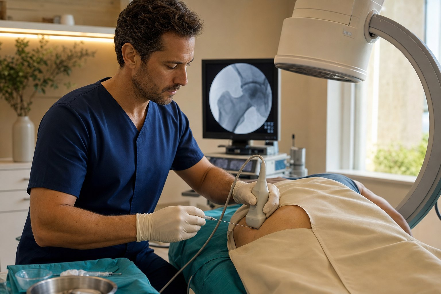
The hip is the joint that best illustrates why a modern regenerative practice invests in both a high-resolution ultrasound machine and a C-arm fluoroscope. Ultrasound is real-time sound imaging — it excels at showing tendon fibers, ligament architecture, the acetabular labrum, capsular thickness, peripheral nerves, and a needle moving through all of them in real time. For the hip, ultrasound is the imaging tool for the labrum, the capsule, the medial and posterior hip tendons, the hamstring origin at the ischial tuberosity, the adductor origin at the pubis, and the femoral and sciatic nerves. Cadaveric and clinical studies of blind hip joint injection show accuracy in the range of 67 to 88 percent depending on operator experience, while ultrasound-guided intra-articular hip injection is accurate approximately 97 percent of the time (Journal of Hip Preservation Surgery accuracy review). At a joint this deep, that difference matters clinically — a missed injection is not just a wasted visit, it is a false-negative diagnostic test and a delay of the correct next step.
Fluoroscopy is real-time X-ray. In the hip it plays a larger role than at the wrist or elbow for two specific reasons. First, intra-articular confirmation of the femoroacetabular joint in advanced osteoarthritis, when the capsule is contracted and osteophytes have distorted the acetabular rim, is often done under ultrasound with fluoroscopic arthrographic confirmation using a small amount of iodinated contrast to prove the needle tip is inside the joint capsule. Second — and this is the target that separates a modern regenerative hip practice from an older one — the intraosseous compartment of the femoral head and acetabulum is a bony target that ultrasound cannot see. Injecting bone marrow concentrate into the subchondral bone requires fluoroscopy and a specific bone-access technique. This is the Hernigou technique, and it is one of the highest-value targets in modern regenerative orthopedics.
Here is the safety caveat that separates a safe hip procedure from an unsafe one: the femoral neurovascular bundle sits directly anterior to the hip joint capsule. Any anterior approach to the femoroacetabular joint puts the femoral nerve, femoral artery, and femoral vein at risk. Ultrasound is not optional for anterior hip work — it is what allows the operator to identify each vascular and neural structure in real time and steer the needle safely past them into the joint.
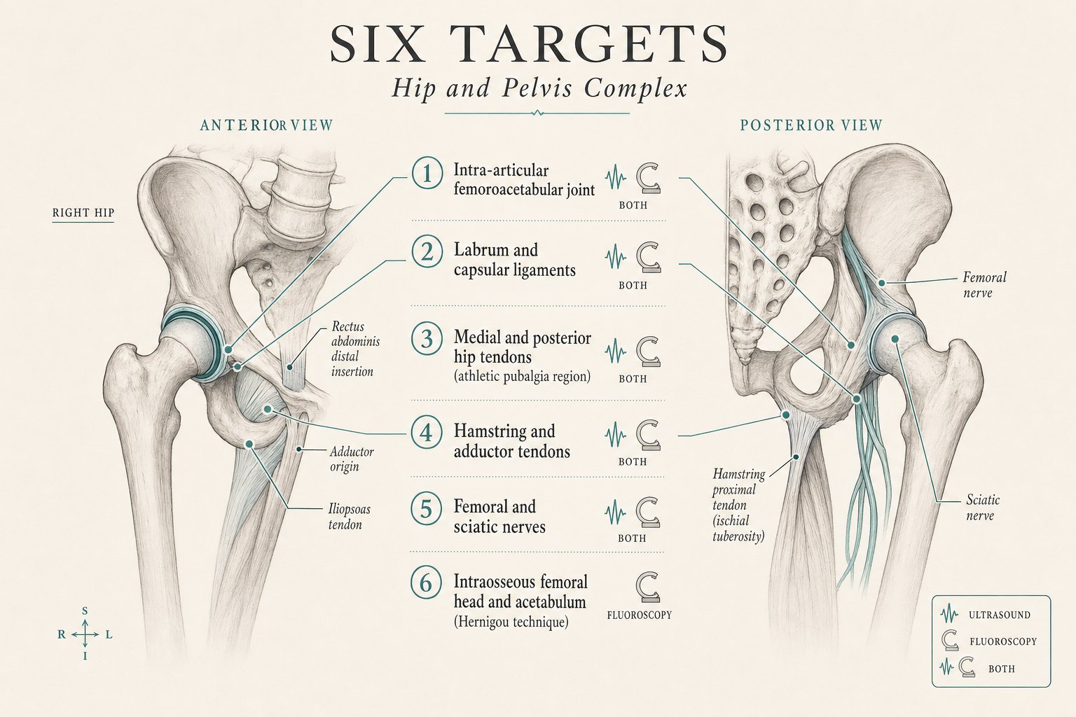
The six sections of the hip ensemble
What follows is a section-by-section walk through each structure I target in a comprehensive regenerative hip procedure. For each one I describe what the structure does when it is healthy, how it manifests as pain when it is not, and how it is targeted — including which imaging tool is right for that specific target.
1. The intra-articular femoroacetabular joint — the deep ball and socket
What it does: The femoroacetabular joint is the articulation between the femoral head and the acetabulum of the pelvis. It is a ball-and-socket synovial joint covered in hyaline cartilage and lined by a synovial membrane, and it bears three to five times body weight during single-leg stance and up to eight times body weight during a sprint. It is one of the most common sites of primary osteoarthritis in the body, and hip osteoarthritis is a leading cause of disability in adults over sixty.
How it manifests as pain: Hip osteoarthritis classically presents as groin pain with a C-shaped grip (the “C sign” where patients cup the greater trochanter and the groin between thumb and fingers), pain with rotation of the hip (getting in and out of a car, putting on socks), pain with weight-bearing rotation, morning stiffness that improves within an hour of activity, and loss of internal rotation on physical exam. It can refer to the buttock, the anterior thigh, and even the medial knee — and patients with true hip pathology are commonly treated for months for the wrong joint before the correct diagnosis is made.
How we target it: With an ultrasound-guided intra-articular femoroacetabular injection, with fluoroscopic arthrographic confirmation added when the joint is severely constrained or when advanced disease makes ultrasound-only confirmation uncertain. The evidence base for hip osteoarthritis PRP has matured substantially. A three-arm randomized controlled trial of 105 patients comparing PRP, hyaluronic acid, and combined PRP+HA found the PRP-containing groups significantly superior at six months on WOMAC (p=0.041) and Lequesne index (p=0.001) (Nouri 2022 RCT). A 111-patient randomized trial found PRP superior to hyaluronic acid at six months with mean VAS pain of 21 versus 44 (p<0.0005) (Dallari 2016 RCT). An 80-patient randomized trial found both PRP and HA effective at twelve months with similar magnitude of benefit (Battaglia RCT). Two systematic reviews conclude that image-guided PRP for hip osteoarthritis is a favorable, safe intervention with a durable therapeutic effect through six to twelve months (Systematic review 2024, Systematic review 2019). The technical takeaway is that this joint requires image guidance every time. It is one of the marquee applications of regenerative medicine at the hip.
2. The acetabular labrum and capsular ligaments — the seal of the joint
What it does: The acetabular labrum is a ring of fibrocartilage that deepens the acetabular socket and creates a suction seal around the femoral head. That seal contributes to load distribution, joint stability, and the maintenance of intra-articular fluid pressure that keeps cartilage nourished. The capsular ligaments — the iliofemoral, pubofemoral, and ischiofemoral bands — are among the strongest ligaments in the body and provide the passive constraint that lets the hip carry load through end-range motion. Labral tears are extraordinarily common in patients with femoroacetabular impingement (FAI), in soccer and hockey athletes, and in adults with dysplastic hip morphology.
How they manifest as pain: Labral tears present as mechanical hip pain with catching, clicking, or a snapping sensation at end-range flexion and internal rotation, positive impingement (FADIR) and log-roll tests on exam, and a distinctive pain pattern that includes groin pain, anterior hip pain with prolonged sitting (the “deep chair sign”), and difficulty with pivoting activities. Capsular laxity from repetitive rotational injury (dancers, gymnasts, martial artists, throwing athletes) presents as microinstability — a vague sense of the hip giving way or feeling “loose,” with a positive prone external rotation test and pain with end-range extension.
How we target them: With ultrasound-guided injection into the labrum and along the capsular ligaments, with the injection often combined with intra-articular hip joint injection. I want to be transparent about the state of the evidence here. Case series of ultrasound-guided PRP for non-surgical partial labral tears have documented meaningful improvement in Harris Hip Score and VAS pain at two, six, and eight weeks (Regenerative Orthopedics and Sports Medicine outcomes), and the technique is increasingly used at high-volume regenerative practices for stage 1-2 labral pathology. However, when PRP has been studied as an intraoperative augmentation to hip arthroscopy for labral repair — the surgical setting — randomized trials have not shown a benefit above the surgical repair itself (Surgical augmentation systematic review). The honest interpretation: for the non-surgical patient with a partial labral tear and reproducible pain who wants to avoid surgery, an image-guided regenerative injection is a legitimate emerging option with early promising data. For the patient who is already a surgical candidate for labral repair, adding PRP to the surgery is not supported by current evidence. The imaging tool is ultrasound, occasionally with fluoroscopic arthrographic confirmation when the joint capsule needs to be entered simultaneously.
3. The medial and posterior hip tendons — athletic pubalgia
What they do: The pubic symphysis is the fibrocartilaginous joint at the anterior midline of the pelvis where the two pubic bones meet. It is the anchor for a remarkable convergence of muscles: the rectus abdominis inserts from above, the adductor longus and adductor brevis originate from below, and the conjoint tendon and posterior inguinal wall complete the network. Under repetitive pivoting, cutting, and kicking loads — the exact loads of soccer, hockey, and lacrosse — this network can break down in a pattern called athletic pubalgia (historically “sports hernia,” though there is no true hernia). The lesion is typically a tear or degeneration of the distal rectus abdominis, the adductor origin, or the conjoint tendon.
How they manifest as pain: Athletic pubalgia presents as chronic lower abdominal pain, groin pain, or medial thigh pain that is provoked by sport-specific movements — kicking, sprinting with change of direction, sit-ups. It is worse with resisted hip adduction and resisted abdominal contraction on exam, and it is a diagnosis of pattern rather than of a single physical finding. It is one of the leading causes of lost playing time in male professional soccer.
How we target them: With ultrasound-guided injection into the specific tendon origin implicated on exam and imaging — most commonly the adductor longus origin and the rectus abdominis distal insertion — with the option of adding hydrodissection along the adjacent iliopsoas tendon. The regenerative evidence for athletic pubalgia is still developing. A published case of a seventeen-year-old soccer player with unilateral adductor tendinopathy who received ultrasound-guided PRP into the adductor complex achieved improvement exceeding the minimum clinically important difference on validated outcome scores and returned to sport (Adductor PRP case, PMC11221341), and case reports from academic sports medicine practices have documented resolution of symptoms and return to competition with image-guided PRP into the adductor and rectus abdominis complex (Wiley PM&R lacrosse case). A 2026 narrative review positions image-guided PRP as a legitimate conservative option in the treatment algorithm for athletic pubalgia, particularly for adductor-predominant patterns, before surgical repair is considered (Narrative review, 2026). This is an area where I set expectations honestly: the evidence is limited but growing, patient selection matters enormously, and the injection is one part of a rehabilitation strategy that must address the underlying core-and-hip loading imbalance.
4. The hamstring and adductor tendons — proximal tendinopathy and tears
What they do: The proximal hamstring tendon is the shared origin of the biceps femoris long head, semimembranosus, and semitendinosus muscles at the ischial tuberosity — the bony prominence you sit on. The adductor tendons originate along the inferior pubic ramus and provide the primary hip adduction and secondary hip flexion moment. Both are load-bearing tendons that fail in a classic pattern of chronic tendinopathy in runners, dancers, cyclists, and equestrians, and can also fail as partial or complete tears from a single high-energy eccentric event (waterskiing, hurdling, a slip on ice with the leg flexed).
How they manifest as pain: Proximal hamstring tendinopathy presents as deep buttock pain at the ischial tuberosity, worse with prolonged sitting (especially on a hard chair), worse with initial acceleration in running or with lunging, tenderness directly on the ischial tuberosity, and pain with resisted knee flexion in end-range hip flexion. Adductor tendinopathy presents as medial groin and thigh pain worse with pivoting and cutting, tenderness at the pubic origin, and pain with resisted hip adduction. Complete tears present as sudden loss of function with a palpable defect and a distal “bunching” bruise.
How we target them: With ultrasound-guided injection of PRP into the tendon body and pathologic pes anserinus or ischial tuberosity origin. The evidence base for image-guided PRP for proximal hamstring tendinopathy has developed over the past decade and supports its use in appropriately selected patients as a durable alternative to prolonged conservative management. The evidence for image-guided treatment of partial and low-grade tears is meaningful and growing. For high-grade complete tears with functional deficit, surgical repair is often the correct answer, and I say so clearly when that is the appropriate recommendation. The imaging tool is ultrasound. Fluoroscopy cannot see these tendons.
5. The femoral and sciatic nerves — regional anesthesia and entrapment
What they do: The femoral nerve is the largest branch of the lumbar plexus and supplies the quadriceps muscles and sensation to the anterior thigh. It runs beneath the inguinal ligament, immediately lateral to the femoral artery, and is a common site for regional anesthesia during knee and anterior thigh surgery. The sciatic nerve is the largest peripheral nerve in the body and exits the pelvis beneath the piriformis muscle before running down the posterior thigh. It is a common site for regional anesthesia for below-knee surgery, and it is the target of deep gluteal syndrome (formerly “piriformis syndrome”) when it becomes entrapped by the piriformis, obturator internus, or hamstring origin.
How they manifest as pain: Deep gluteal syndrome presents as deep buttock pain worse with prolonged sitting, sciatic-distribution pain into the posterior thigh (occasionally to the foot), tenderness deep to the piriformis on exam, and a positive seated piriformis test. Femoral neuropathy is less commonly a chronic pain syndrome and more commonly a post-surgical or entrapment issue in specific settings.
How we target them: With ultrasound-guided perineural block for regional anesthesia, or ultrasound-guided hydrodissection with five percent dextrose (D5W) or platelet lysate for entrapment neuropathies. The hydrodissection principles that apply to the median nerve at the carpal tunnel and the ulnar nerve at the cubital tunnel apply here as well: a fluid bolus is used to gently separate the nerve from the surrounding fascia, muscle, or scar tissue that is compressing it. Ultrasound is essential; both the femoral and sciatic nerves are invisible to fluoroscopy, and the femoral artery sits immediately medial to the femoral nerve at the level where any perineural injection would be performed. Real-time visualization is the standard.
6. The intraosseous femoral head and acetabulum — the Hernigou technique
What it does: The subchondral bone marrow of the femoral head and the acetabulum sits just beneath the articular cartilage of the femoroacetabular joint. It is a metabolically active zone that contains a native population of mesenchymal stem cells, and it is where the earliest changes of advanced osteoarthritis and the entirety of stage 1-2 avascular necrosis (AVN) of the femoral head take place. Bone marrow lesions (edema patterns on MRI in the subchondral zone) correlate strongly with pain and progression in hip osteoarthritis and are one of the most reliable radiographic predictors of who will progress toward total hip arthroplasty.
How it manifests as pain: Intraosseous pathology presents as a specific pain pattern — deep, poorly-localizable groin pain that is worse with weight-bearing and often worse at rest at night in advanced disease, poorly responsive to intra-articular injection because the pain generator is in the bone rather than in the joint capsule, and correlated on MRI with subchondral bone marrow edema, cystic change, or, in the case of AVN, a crescent sign or subchondral collapse. It is the pain pattern that distinguishes patients whose osteoarthritis will respond to an intra-articular injection alone from patients whose disease has progressed to the bone and requires an intraosseous approach.
How we target it: With a fluoroscopically-guided intraosseous injection of bone marrow aspirate concentrate (BMAC) into the femoral head and, when appropriate, the acetabulum — the Hernigou technique. This is the target that most distinguishes a modern regenerative hip practice from a conventional one, and the evidence has become genuinely compelling. The original technique was developed by Philippe Hernigou for stage I and II femoral head avascular necrosis, and his long-term follow-up demonstrated that in a cohort of 145 hips treated with concentrated bone marrow, only 9 required total hip arthroplasty at five- to ten-year follow-up — a striking result for a disease whose natural history without intervention is progression to collapse and arthroplasty (Hernigou original series, PMC3272721). A modified core decompression technique combining core decompression with intraosseous BMAC in thirty-two hips with early-stage AVN documented no progression except in four patients with bilateral disease (Modified core decompression 2024). A meta-analysis of BMAC for femoral head AVN found improvement in early-stage disease but limited efficacy in post-collapse disease — a critical staging distinction (Meta-analysis BMAC AVN, PMC7919970). For advanced osteoarthritis with bone marrow lesions, the Regenexx group has published fifteen-year outcomes from a 217-patient controlled study demonstrating that patients treated with intra-articular and intraosseous BMAC had a total hip arthroplasty rate at fifteen years of 16.1 percent compared to 40.1 percent in a conservative-care comparison group, with statistically superior Kaplan-Meier survival (Regenexx 15-year outcomes). This is a target that requires fluoroscopy for bone access, requires appropriate patient staging (early-stage AVN and osteoarthritis with bone marrow lesions are the ideal indications; post-collapse AVN and end-stage arthritis are surgical decisions), and requires the technical experience to safely access the bone. It is one of the most impactful interventions available in modern regenerative orthopedics.
How the conductor puts the hip ensemble together
A comprehensive regenerative hip procedure at Pravida does not treat all six structures on every patient. The point of the physical exam, the imaging review, and the diagnostic history is to determine which sections of the ensemble are actually out of tune for you. Some patients have a single-structure problem — an isolated osteoarthritis of the femoroacetabular joint, an isolated proximal hamstring tendinopathy, an isolated deep gluteal syndrome — and a single well-placed injection is exactly the right answer. Other patients have combined patterns (the most common in adults over fifty is intra-articular osteoarthritis plus subchondral bone marrow lesions in the femoral head; in soccer athletes it is labral pathology plus adductor and rectus abdominis pubalgia; in runners it is hamstring tendinopathy plus deep gluteal irritation), and treating just one of them is precisely why the last two shots wore off in weeks.
What a world-class hip procedure looks like on the day it happens: a physical exam and imaging review that identifies the specific structures in play, a written plan for which structures will be treated, a procedure suite set up with both a high-resolution ultrasound machine and a C-arm fluoroscope, and a sequence of small, precise injections — each one confirmed on real-time imaging before medication is delivered — that treats each identified section of the ensemble with the tool that shows it best. Ultrasound is used for the labrum, capsule, medial and posterior hip tendons, hamstring and adductor origins, and peripheral nerves. Ultrasound with fluoroscopic arthrographic confirmation is used for the femoroacetabular joint in advanced disease. Fluoroscopy is used for intraosseous work at the femoral head and acetabulum. Most hip procedures are outpatient and take under ninety minutes. Most patients drive themselves home the same day, though weight-bearing precautions apply to the intraosseous protocol.
The choice of biologic — platelet-rich plasma (typically leukocyte-poor for tendon, ligament, and labral work), bone marrow concentrate for higher-grade partial ligament injury, advanced joint disease, or intraosseous work, or five percent dextrose for nerve hydrodissection — is guided by the tissue being treated and by the patient’s overall regenerative plan. The CartiNova program is our organized framework for making those choices in a way that is transparent, evidence-informed, and personalized to the individual patient.
What most hip patients get wrong before they see us
- They accept a blind hip shot as the standard first step. Blind injection at the hip is accurate 67-88 percent of the time even in experienced hands; ultrasound-guided injection is accurate 97 percent of the time. A missed injection is not a failed treatment — it is a false-negative diagnostic test that delays the correct next step. Image-guided technique is the standard.
- They treat the wrong joint. Groin pain, anterior thigh pain, and medial knee pain are commonly hip pathology, and patients with true hip disease are often treated for months for the wrong joint. A comprehensive hip evaluation looks at the femoroacetabular joint, the labrum, the tendon insertions, the nerves, and the intraosseous compartment before a diagnosis is made.
- They ignore the bone. In advanced osteoarthritis, the intra-articular injection often wears off in weeks because the pain generator is in the subchondral bone, not in the joint capsule. Bone marrow lesions on MRI and stage 1-2 AVN of the femoral head are the specific indications for the Hernigou intraosseous technique — and this is the intervention that changes the disease trajectory when it is done correctly.
- They confuse a surgical decision with an injection decision. Stage 3-4 hip osteoarthritis with bone-on-bone joint space loss is a total hip arthroplasty decision. Post-collapse AVN with femoral head deformity is a surgical decision. Complete adductor or hamstring rupture in a young athlete is often a repair decision. A good regenerative practice will tell you clearly when injection is the right answer and when it is not.
- They expect regeneration without rehabilitation. A well-placed biologic creates a window in which targeted hip strengthening, gluteal activation, and progressive loading exercises can rebuild the tissue and restore function. The injection and the rehab are one intervention, not two.
The honest limits, and where a good clinician still matters
- The six-structure approach is designed for patients with chronic or subacute hip and groin pain that has not fully resolved with conservative care. It is not first-line treatment for advanced (stage 3-4) hip osteoarthritis with bone-on-bone joint space loss, post-collapse femoral head avascular necrosis, complete proximal hamstring or adductor rupture in a young athlete, acute traumatic dislocation, septic arthritis, or red-flag presentations (fever, unexplained weight loss, night pain unresponsive to positional change).
- The regenerative evidence base is strongest for image-guided intra-articular PRP in hip osteoarthritis and for the Hernigou intraosseous technique in early-stage AVN and advanced osteoarthritis with bone marrow lesions. It is meaningful and developing for non-surgical labral pathology, proximal hamstring tendinopathy, and adductor tendinopathy. It is limited but promising for athletic pubalgia. I discuss the strength of the evidence for the specific target with every patient before we proceed.
- Any anterior hip procedure requires ultrasound. If a clinician offers a fluoroscopically-guided-only injection at the anterior femoroacetabular joint or along the anterior capsule, they are potentially working within millimeters of the femoral neurovascular bundle without visualizing it. Real-time ultrasound is the standard.
- Regenerative outcomes depend on the underlying tissue, the biologic used, the technical precision of delivery, and the rehabilitation that follows. A biologic delivered blindly, or into the wrong structure, is not regenerative — it is just an injection.
- If you have new onset of severe hip pain with fever, sudden inability to bear weight after minor trauma, pain accompanied by new neurologic deficit, or new hip pain in the setting of active cancer, please contact your physician or an emergency department today. Those are not situations for an elective regenerative procedure.
Ready to have your specific hip pattern mapped?
If you are an Atlanta-area patient with chronic hip, groin, or buttock pain that has not fully responded to prior injections or conservative care — or you are researching alternatives to total hip arthroplasty, core decompression, or labral repair before you commit to surgery — we would rather see you in person. A consultation at Pravida Health includes a physical exam, an imaging review, and, where appropriate, a comprehensive regenerative plan tailored to the specific sections of your hip ensemble that are out of tune.
Book a consultationKey sources referenced in this article
- Nouri F, et al. Comparison of PRP, hyaluronic acid, and combined PRP+HA for hip osteoarthritis: three-arm randomized controlled trial. PubMed 36096771. PRP-containing groups significantly superior at six months on WOMAC (p=0.041) and Lequesne (p=0.001).
- Dallari D, et al. PRP versus hyaluronic acid for hip osteoarthritis: 111-patient randomized trial. PubMed 26797697. PRP VAS 21 versus HA 44 at six months (p<0.0005).
- Battaglia M, et al. PRP versus hyaluronic acid for hip osteoarthritis: 80-patient randomized trial. PMC5738493. Both effective at twelve months with similar magnitude of benefit.
- Systematic review of PRP for hip osteoarthritis, 2024. PMC11578636. Favorable therapeutic effect and safety profile through six to twelve months.
- Systematic review of PRP for hip osteoarthritis, 2019. PMC6444309. Consistent short-to-medium-term benefit; image-guided technique required.
- Ultrasound-guided versus blind hip joint injection accuracy. Journal of Hip Preservation Surgery, Oxford Academic. Ultrasound guidance 97 percent accurate versus 67-88 percent for blind technique.
- Ultrasound-guided PRP for hip labral tears: case series. Regenerative Orthopedics and Sports Medicine outcomes. Harris Hip Score and VAS improvement at 2, 6, and 8 weeks in non-surgical partial labral tears.
- Systematic review of PRP augmentation in hip arthroscopy for labral repair. PMC8460156. Intraoperative PRP augmentation does not improve outcomes above surgical labral repair alone.
- Ultrasound-guided PRP for adductor tendinopathy in a young soccer player: case with MCID exceeded. PMC11221341.
- PRP for athletic pubalgia in a lacrosse player: case report. Wiley PM&R, 2014. Image-guided PRP into the adductor and rectus abdominis complex.
- Narrative review of conservative and regenerative treatment of athletic pubalgia, 2026. PMC12832563. Image-guided PRP as a conservative option in the treatment algorithm before surgical repair.
- Hernigou P, et al. Bone marrow concentrate for stage I and II femoral head avascular necrosis: long-term follow-up. PMC3272721. Only 9 of 145 hips required total hip arthroplasty at 5- to 10-year follow-up.
- Modified core decompression with intraosseous BMAC for early-stage AVN, 2024. PubMed 39398835. No progression except in four patients with bilateral disease across 32 hips.
- Meta-analysis of BMAC for femoral head avascular necrosis. PMC7919970. BMAC effective in early-stage AVN; limited efficacy in post-collapse disease.
- Regenexx 15-year controlled outcomes: intraosseous and intra-articular BMAC for hip osteoarthritis. 15-year outcome study, 217 patients. Total hip arthroplasty rate 16.1 percent BMAC vs. 40.1 percent conservative at 15 years; superior Kaplan-Meier survival.