
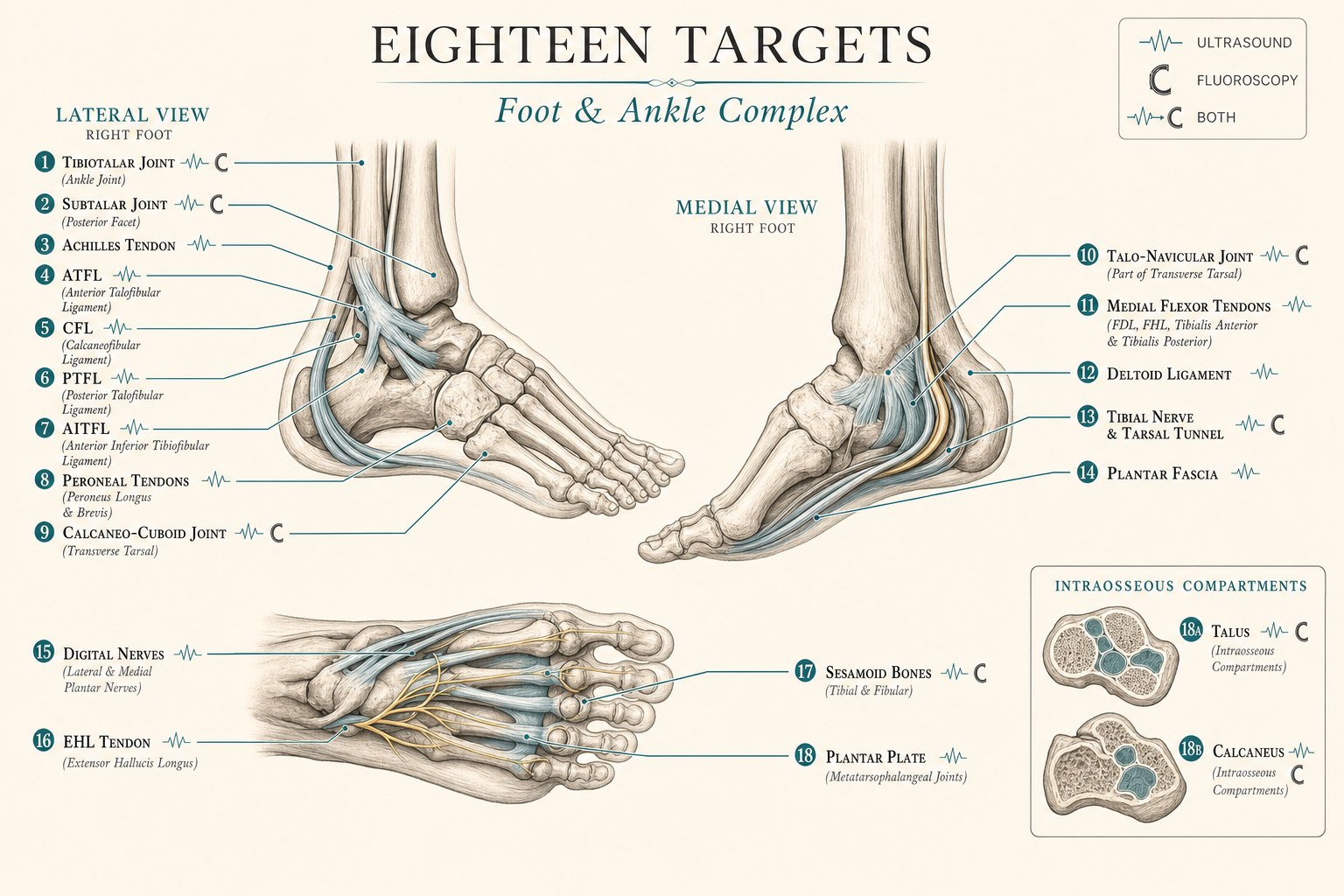
The foot and ankle as an eleven-part symphony
The foot and ankle carry the entire weight of the body across every step of a lifetime — roughly seven thousand steps a day for the average American adult, and easily twice that for an active one. Twenty-six bones, thirty-three joints, more than a hundred ligaments, and a densely packed collection of tendons, nerves, and fascia are asked to translate the ground into forward motion, catch the body on uneven terrain, and absorb load equivalent to three to five times body weight with every running stride. It is one of the most mechanically complex regions in the body, and one of the most under-treated: the majority of patients with chronic foot or ankle pain arrive having been offered essentially two options — a blind steroid injection or a surgical opinion. Regenerative interventional medicine sits deliberately between those two poles.
This is the seventh article in a series on how a master regenerative procedure actually works, region by region. We started with the five-structure ensemble of the lumbar spine, walked through the fourteen-structure symphony of the shoulder, mapped the seven-structure ensemble of the elbow, detailed the ten-structure precision of the wrist and hand, traced the six-structure architecture of the hip and pelvis, and mapped the nine-structure symphony of the knee. The foot and ankle bring a distinctive challenge: they present with an unusually wide range of pathology types — joint, tendon, ligament, fascia, nerve, and bone marrow — often in the same patient. A runner with plantar fasciitis frequently also has posterior tibial tendinopathy, a low-grade ATFL sprain from a prior ankle turn, and a small subtalar joint arthritis that has developed as compensation. Treating one and ignoring the others is exactly why so many patients feel that their “heel injection” wore off in weeks.
What follows is the conductor’s map for the foot and ankle: every structure I target in a comprehensive regenerative foot and ankle procedure, why each one matters, how it produces pain when it is out of tune, and which imaging tool — ultrasound, fluoroscopy, or both — is right for that section. I want to be transparent up front about one point that will run through this article: the evidence base for regenerative medicine in the foot and ankle is not uniform. It is excellent for plantar fasciitis. It is promising and case-series level for partial ATFL and lateral ankle ligament injuries. It is emerging for peroneal tendinopathy and for talar osteochondral lesions with BMAC. It is controversial and mostly negative for tibiotalar joint osteoarthritis and for Achilles tendinopathy, and I will say so plainly rather than sell around it.
Why two imaging tools, and why the foot and ankle need both
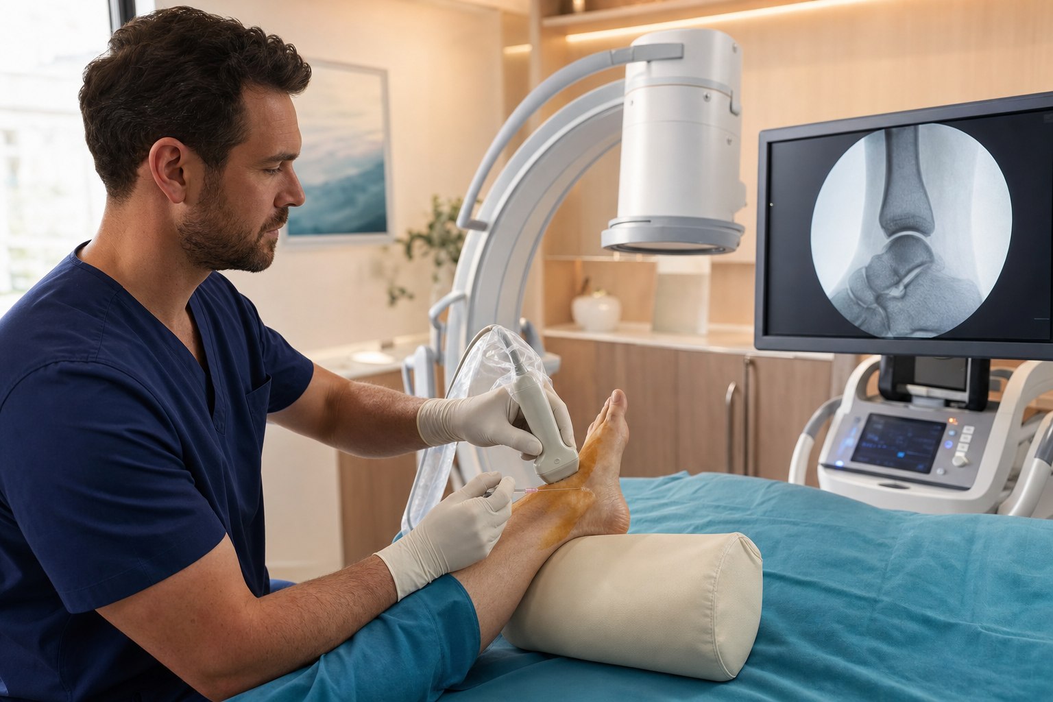
The foot and ankle contain some of the most superficial structures in the body (the peroneal tendons, the ATFL, the plantar fascia, the tibial nerve at the tarsal tunnel) sitting immediately next to some of the deepest and bone-shrouded targets in the body (the tibiotalar joint, the subtalar posterior facet, and the subchondral bone of the talus and calcaneus). No single imaging tool sees both well. Ultrasound is real-time sound imaging — it excels at showing tendon fiber pattern, ligament architecture, fascia thickness, small joint capsules, peripheral nerves, and a needle moving through all of them in real time. For the foot and ankle, ultrasound is the imaging tool for the Achilles tendon, plantar fascia, ATFL/CFL/PTFL/AITFL, deltoid ligament, peroneal tendon body, posterior tibial and flexor tendons, EHL tendon, sesamoids, plantar plate, tibial nerve at the tarsal tunnel, digital nerves, and the small joints of the midfoot when the operator has the skill for them. Ultrasound sees soft tissue; it cannot see through bone.
Fluoroscopy is real-time X-ray, and it is the tool for the deep, bone-shrouded targets. In the foot and ankle it plays a specific and important role in four situations. The first is intra-articular access to the tibiotalar and subtalar joints in patients with joint-space narrowing, where an ultrasound-only approach can be blocked by osteophytes or a scarred capsule and fluoroscopically-confirmed contrast diffusion demonstrates that the biologic has actually reached the joint. The second is the retromalleolar peroneal tendon sheath, where fluoroscopy with a small amount of iodinated contrast confirms sheath (rather than intratendinous) delivery in patients whose sheath has become fibrosed. The third is the intraosseous compartment of the talus and calcaneus for advanced bone marrow lesions and osteochondral lesions of the talus. Bone is invisible to ultrasound; fluoroscopy is what lets the operator place the needle tip accurately in the subchondral bone. The fourth is any procedure where iodinated contrast imaging is needed to verify diffusion pattern and rule out inadvertent tendon injection.
Here is the safety principle that separates a modern foot and ankle procedure from an older one: image guidance is the standard for every target, superficial or deep. The tibial nerve runs immediately deep to the medial flexor tendons at the tarsal tunnel. The peroneal tendons sit immediately posterior to the fibula and lateral to the sural nerve. The saphenous nerve accompanies the great saphenous vein anterior to the medial malleolus. The digital nerves sit immediately alongside the vessels between the metatarsal heads. A needle placed blindly into any of these zones risks nerve or vascular injury. Image guidance is not a luxury — it is what makes the procedure safe as well as accurate.
The eleven sections of the foot and ankle ensemble
What follows is a section-by-section walk through each structure or group of structures I target in a comprehensive regenerative foot and ankle procedure. For each one I describe what the structure does when it is healthy, how it manifests as pain when it is not, and how it is targeted — including which imaging tool is right for that specific target, and where the evidence sits in September 2026.
1. The tibiotalar joint — the main ankle joint
What it does: The tibiotalar joint is the true ankle joint — the articulation between the distal tibia and fibula superiorly, forming the mortise, and the trochlea of the talus inferiorly. It is a modified hinge joint responsible for dorsiflexion and plantarflexion, and it carries three to five times body weight during walking and up to thirteen times body weight during running. Its articular cartilage is thin relative to the knee (roughly 1.3 to 1.7 mm on average versus 2.4 to 2.5 mm) which explains part of why the ankle is more resistant to primary osteoarthritis than the knee but far more susceptible to post-traumatic osteoarthritis after a serious sprain or fracture.
How it manifests as pain: Tibiotalar osteoarthritis or synovitis presents as anterior ankle pain worse with weight-bearing, morning stiffness that eases within thirty to sixty minutes of activity, loss of full dorsiflexion, anterior joint-line tenderness, and, in advanced disease, effusion and a bone-on-bone end feel. Roughly seventy to eighty percent of ankle osteoarthritis is post-traumatic in origin, distinguishing it clinically and prognostically from primary knee osteoarthritis.
How we target it: With a fluoroscopically- or ultrasound-guided anterior intra-articular tibiotalar injection. I need to be transparent here because this is one of the places where the regenerative evidence diverges sharply from the knee. The best-designed randomized controlled trial of intra-articular PRP for ankle osteoarthritis is Paget’s 2021 JAMA trial — a rigorous 100-patient double-blind study comparing two ultrasound-guided PRP injections to two saline injections in patients with tibiotalar osteoarthritis. At twenty-six weeks, the primary endpoint (AOFAS score) showed no statistically significant difference between PRP and saline (Paget 2021 JAMA, PMC8548954). The fifty-two-week follow-up continued to show no meaningful difference (Paget 52-week follow-up, PMC10394962). Smaller before-and-after case series have shown short-term symptomatic improvement (Case-series before-after, PMC10197236), but the highest-quality evidence is that intra-articular PRP alone is not a proven treatment for isolated tibiotalar osteoarthritis. This is different from the knee, where the intra-articular PRP evidence is genuinely strong, and I say so directly with every patient. Where an isolated tibiotalar injection does have a legitimate role is (a) for diagnostic and short-term symptom relief when the source of the pain is unclear, (b) as one component of a comprehensive procedure when there is coexisting soft-tissue and intraosseous pathology, and (c) for anterior ankle synovitis or capsulitis (rather than for advanced bony osteoarthritis).
2. The subtalar joint — the hindfoot workhorse
What it does: The subtalar joint sits immediately beneath the talus, between the talus above and the calcaneus below. It has three articular facets (anterior, middle, posterior), and functionally it is the joint that translates dorsiflexion and plantarflexion at the ankle into the eversion and inversion that let the foot adapt to uneven ground. The posterior facet is the largest and most clinically important; it is the target when we talk about “the subtalar injection.” Subtalar arthritis is a common consequence of calcaneal fractures, of severe lateral ankle sprains that heal with instability, of chronic peroneal tendinopathy, and of adult acquired flatfoot deformity.
How it manifests as pain: Subtalar pathology presents as pain deep in the sinus tarsi region (immediately anterior and inferior to the lateral malleolus), pain with walking on uneven surfaces or on grass, pain with side-to-side motion of the hindfoot on exam, and tenderness on palpation of the sinus tarsi. It is one of the most under-diagnosed sources of “lateral ankle pain,” and patients are frequently misdiagnosed with a chronic ATFL problem when their pain generator is actually the subtalar joint.
How we target it: With a fluoroscopically-guided posterior subtalar joint injection, or, in patients with an accessible sinus tarsi and preserved joint architecture, an ultrasound-guided lateral sinus tarsi approach. Fluoroscopy is preferred when the subtalar joint space is narrowed on imaging or when previous injection attempts have not clearly reached the joint. Contrast diffusion is used to confirm intra-articular position. Intra-articular PRP for isolated subtalar arthritis has been reported in case series with symptomatic improvement, but the evidence is substantially thinner than for peripheral joints. As with the tibiotalar joint, the subtalar injection is often most valuable as one instrument in a broader regenerative procedure rather than as a standalone intervention.
3. The transverse tarsal joints — talo-navicular and calcaneo-cuboid
What they do: The transverse tarsal joints — the talo-navicular joint medially and the calcaneo-cuboid joint laterally — sit at the boundary between the hindfoot and the midfoot. Together they form the Chopart joint line. They are critical to normal foot mechanics: when the calcaneus everts, the two joint axes align and the midfoot becomes flexible for shock absorption; when the calcaneus inverts, the axes diverge and the midfoot locks into a rigid lever for push-off. Osteoarthritis or ligamentous injury at these joints is a common source of midfoot pain, particularly after inversion injuries, in patients with pes planus, and in patients with a history of adult acquired flatfoot deformity or Charcot arthropathy.
How they manifest as pain: Talo-navicular pain presents as dorsomedial midfoot pain worse with push-off and with prolonged standing, tenderness on palpation of the talo-navicular joint line, and, in advanced disease, a palpable dorsal osteophyte. Calcaneo-cuboid pain presents as lateral midfoot pain worse with cutting, twisting, and pivoting activities, tenderness on palpation of the lateral joint line just distal to the anterior process of the calcaneus, and, occasionally, referred pain to the base of the fifth metatarsal.
How we target them: With ultrasound-guided intra-articular injection of the specific joint of pathology. Both joints are readily accessed under ultrasound in most patients with mild-to-moderate arthritis, and image guidance is essential because the neighboring joints and dorsal cutaneous nerves are close and easily misinjected. Evidence for regenerative therapy at these specific joints is limited to case series and extrapolation from the small-joint osteoarthritis literature; the more common indication in my practice is targeted intra-articular corticosteroid for acute inflammation with a followup PRP course for maintenance in appropriate patients.
4. The Achilles tendon — the ensemble’s most-discussed and most-mixed target
What it does: The Achilles tendon is the largest and strongest tendon in the body, formed by the confluence of the gastrocnemius and soleus muscles and inserting onto the posterior calcaneus. It transmits the entire plantarflexion force of the calf, and it carries load equivalent to twelve times body weight during running. Achilles tendinopathy is classically divided into midportion (roughly two to six centimeters proximal to the insertion) and insertional (immediately at the calcaneus), with different clinical courses and different treatment responses.
How it manifests as pain: Midportion Achilles tendinopathy presents as posterior lower-leg pain, morning stiffness of the tendon that improves with a few minutes of walking, fusiform thickening of the tendon two to six centimeters above the calcaneus, and pain with resisted plantarflexion. Insertional Achilles tendinopathy presents as pain right at the posterior calcaneus, often with a Haglund’s deformity or an insertional bone spur, and is characteristically more difficult to treat than midportion disease.
How we target it: With an ultrasound-guided PRP injection into the pathologic tendon, combined with a structured heavy-slow-resistance loading program. I need to be more transparent about this target than any other in this article. The best-designed randomized controlled trials of PRP for Achilles tendinopathy are largely negative. The 2016 Krogh RCT found no benefit of PRP versus saline for chronic Achilles tendinopathy (Krogh 2016, PubMed 27257167). The 2020 Boesen trial similarly found no PRP benefit versus placebo (Boesen 2020, PubMed 32485112). The 2019 PATH-2 trial of PRP for acute Achilles rupture also found no benefit (PATH-2, BJSM). A 2024 meta-analysis by Kraychete concluded that PRP is not effective and should not be used for chronic Achilles tendinopathy (Kraychete 2024, PubMed 39745256). Set against those, some positive prospective case series and observational studies do exist (Positive case series, PMC11952083), and technique and patient selection remain areas of active investigation. The honest position is this: image-guided PRP for chronic Achilles tendinopathy is not a first-line intervention, and it is not a reliably successful one. What I offer patients with chronic Achilles tendinopathy that has failed a proper heavy-slow-resistance loading program is a conversation about that evidence, and, in carefully selected cases, an image-guided PRP procedure done in conjunction with (not as a substitute for) a structured loading rehabilitation. Insertional disease with a bone spur or a Haglund’s deformity frequently needs a surgical consultation rather than an injection.
5. The plantar fascia — where the regenerative evidence is genuinely strong
What it does: The plantar fascia is a dense fibrous band that originates on the medial calcaneal tubercle and inserts onto the base of each proximal phalanx via five slips. It functions as the roof of the plantar arch, absorbing load during heel strike and midstance, and it acts as a windlass mechanism during push-off — tightening as the toes dorsiflex to elevate the arch. Plantar fasciitis is one of the most common orthopedic complaints in adults; it affects roughly one in ten adults over a lifetime and accounts for over one million clinical visits per year in the United States.
How it manifests as pain: Plantar fasciitis classically presents as sharp medial heel pain worst with the first few steps in the morning or after prolonged sitting, improving with a few minutes of walking, worsening again after prolonged standing or at the end of the day, and tender on palpation of the medial calcaneal tubercle. On ultrasound, the involved plantar fascia is typically thickened beyond four millimeters, hypoechoic in its proximal portion, and sometimes shows a small calcaneal enthesophyte.
How we target it: With an ultrasound-guided PRP injection into the pathologic fascia at the medial calcaneal origin, combined with a structured stretching and load-management program. This is the target in the foot and ankle where the regenerative evidence is unambiguously strong. Hohmann’s 2021 meta-analysis of nine randomized controlled trials found that PRP was superior to corticosteroid injection on VAS at three, six, and twelve months (Hohmann 2021 meta-analysis, SAGE 10.1177/0363546520937293). Bucak’s 2025 trial showed that PRP produced superior Foot Function Index scores at six months, with a clinically meaningful difference exceeding the minimum clinically important difference (Bucak 2025, SAGE 10.1177/10711007251346784). Sherpa’s 2024 systematic review of thirteen trials again showed PRP superior to corticosteroid at mid-term follow-up (Sherpa 2024, PubMed 40913486). Peerbooms’ long-term follow-up at eighteen months documented VAS scores of 2.1 in the PRP group versus 3.6 in the corticosteroid group (Peerbooms long-term, PubMed 30448183). Chew’s trial reported that 84.4 percent of PRP-treated patients achieved a 25 percent or greater improvement in Foot Function Index versus 55.6 percent of corticosteroid-treated patients (Chew, PubMed 31603721). Importantly, PRP avoids the risk of plantar fat pad atrophy that has been documented after repeated steroid injection. Image-guided PRP for plantar fasciitis is, alongside intra-articular PRP for knee osteoarthritis, one of the best-supported interventions we offer.
6. The lateral ankle ligaments — ATFL, CFL, PTFL, and AITFL
What they do: The lateral ankle ligament complex is a group of small stabilizing ligaments that prevent inversion and anterior translation of the talus. The anterior talofibular ligament (ATFL) runs from the anterior fibula to the anterolateral talar neck and is the primary restraint against inversion. It is the most commonly injured ligament in the entire body — roughly one ankle sprain per ten thousand people per day in the United States. The calcaneofibular ligament (CFL) runs from the tip of the fibula to the lateral calcaneus and is the secondary restraint against inversion, injured in about half of severe ankle sprains. The posterior talofibular ligament (PTFL) is a stout deep ligament rarely injured except in complete ankle dislocations. The anterior inferior tibiofibular ligament (AITFL) is the low anterior component of the syndesmosis and is the key ligament injured in a “high ankle sprain,” classically from a dorsiflexion-external-rotation mechanism.
How they manifest as pain: ATFL injury presents as anterolateral ankle pain and tenderness at the anterior fibula, positive anterior drawer, swelling and bruising over the anterior lateral ankle, and, in chronic injury, mechanical instability with cutting and pivoting activities. CFL injury adds tenderness at the lateral calcaneus. AITFL/high ankle sprain presents as anterior distal tibiofibular pain, pain with a squeeze test at the mid-calf, pain with external rotation stress, and a substantially longer recovery arc than a lateral ankle sprain — often three to four times as long.
How we target them: With ultrasound-guided PRP injection into the specific ligament of pathology, together with a structured proprioceptive rehabilitation program. The evidence base for image-guided PRP at the lateral ankle ligaments is genuinely favorable at the case-series level. A published case documented complete ATFL healing after a single ultrasound-guided PRP injection (Complete ATFL healing case, PMC5867359). Supportive protocol and outcomes papers describe PRP-augmented healing of partial ATFL tears (Supportive protocol, PMC9817145, Additional supportive series, PMC12873745). An ultrasound-guided ATFL repair series documented sustained clinical benefit at two-year follow-up (Two-year outcomes, PubMed 41276119, Technique paper, PubMed 39798604). Diagnostic ultrasound assessment of the ATFL is more sensitive than static clinical exam (Ultrasound assessment, PMC6942111). I am transparent about one important caveat: a randomized trial of leukocyte-reduced PRP as an adjunct after Brostrom lateral ligament reconstruction did not show benefit over standard postoperative management (Post-Brostrom PRP negative RCT, PMC10141288). What the evidence supports is image-guided PRP for partial ATFL and lateral ligament injuries in non-surgical patients — not as a routine surgical adjunct.
7. The deltoid ligament — the medial ankle’s stabilizer
What it does: The deltoid ligament complex is the large fan-shaped ligament of the medial ankle, running from the medial malleolus to the talus, calcaneus, and navicular. It has a deep component (primarily the anterior tibiotalar ligament) and a superficial component (tibiocalcaneal, tibiospring, and tibionavicular fibers). Its role is to prevent eversion and lateral translation of the talus in the mortise and to stabilize the medial column of the foot. Isolated deltoid injuries are less common than lateral ankle sprains, but they occur in eversion-external-rotation mechanisms, in high ankle sprains with a rotational component, and in association with adult acquired flatfoot deformity as the deltoid attenuates chronically.
How it manifests as pain: Deltoid ligament injury presents as medial ankle pain and tenderness at the medial malleolus, pain with eversion stress, medial swelling and bruising, and, in chronic injury, medial-side ankle instability. Chronic deltoid attenuation in the setting of stage 2 or stage 3 adult acquired flatfoot deformity presents as progressive medial arch collapse, medial ankle pain, and posterior tibial tendon dysfunction.
How we target it: With an ultrasound-guided PRP injection into the specific area of pathology on the deep or superficial deltoid, with careful attention to the tibialis posterior tendon immediately anterior and inferior. Regenerative evidence for isolated deltoid injury is limited to case reports and small series; the technique and rationale are extrapolated from the lateral ankle ligament literature. Grade 3 deltoid rupture with functional instability and adult acquired flatfoot deformity are typically surgical decisions.
8. The peroneal tendons — laterally overworked, laterally under-treated
What they do: The peroneus longus and peroneus brevis are the two long tendons of the lateral compartment of the leg. They pass behind the lateral malleolus in a shared synovial sheath at the retromalleolar groove, where the peroneus brevis lies anteromedial to the peroneus longus. Peroneus brevis inserts on the base of the fifth metatarsal; peroneus longus wraps under the cuboid and inserts on the base of the first metatarsal and medial cuneiform. Together they evert the foot and provide dynamic stabilization of the lateral ankle. Chronic lateral ankle instability, cavovarus foot morphology, and repetitive lateral-loading activities predispose to peroneal tendinopathy, tenosynovitis, and split tears of the peroneus brevis.
How they manifest as pain: Peroneal tendinopathy presents as chronic lateral ankle and posterior fibular pain, swelling along the peroneal tendon course, tenderness on palpation of the retromalleolar groove and lateral calcaneus, pain with resisted eversion, and, in advanced cases, a snapping or clicking sensation with peroneal subluxation over the lateral malleolus.
How we target them: With an ultrasound-guided PRP injection into the pathologic tendon body or peritendinous sheath, and, in patients with a fibrosed retromalleolar sheath, a fluoroscopically-guided sheath injection to confirm sheath (not intratendinous) delivery. The regenerative evidence for peroneal tendinopathy is at the case-series level. Intratendinous PRP with structured rehabilitation has been reported to produce meaningful improvement in prospective series (Peroneal PRP case series), and dynamic ultrasound of the peroneal tendons has been shown to outperform static MRI for peroneal pathology, making image-guided intervention particularly well-suited to this target (Dynamic ultrasound of peroneal tendons). Split tears with mechanical symptoms and frank peroneal subluxation are surgical decisions rather than injection decisions.
9. The medial tendons and small forefoot structures
What they do: The medial ankle tendons include the tibialis posterior (the primary dynamic stabilizer of the medial arch), the flexor digitorum longus (FDL, flexor of the lateral four toes), and the flexor hallucis longus (FHL, flexor of the great toe and often called “the ballerina’s tendon”). The anterior compartment adds the tibialis anterior (the primary dorsiflexor) and the extensor hallucis longus (EHL, the great-toe extensor). The forefoot adds the sesamoid bones (two small bones embedded within the FHL tendon plantar to the first metatarsal head, absorbing load with push-off) and the plantar plates (fibrocartilaginous structures on the plantar surface of each metatarsophalangeal joint that stabilize the toes and prevent hyperextension). Together this group is asked to power push-off, stabilize the arch, dorsiflex the ankle, and translate load through the forefoot on every step.
How they manifest as pain: Posterior tibial tendinopathy presents as medial ankle and arch pain, arch collapse over time (stage 1-4 adult acquired flatfoot), and pain with resisted inversion — a classic single-heel-rise test failure. FHL tendinopathy classically presents in ballet dancers as posteromedial ankle pain worse with pointing the great toe. Tibialis anterior tendinopathy presents as anterior ankle pain worse with prolonged walking or running, often with a palpable tender nodule at the tendon anterior to the ankle. Sesamoiditis presents as pain plantar to the first metatarsal head, worse with push-off and toe-off. Plantar plate tears present as pain at the plantar aspect of the second, third, or fourth metatarsophalangeal joint, often with a positive drawer sign at that toe and a gradually developing hammer toe deformity.
How we target them: With ultrasound-guided PRP injection into the pathologic tendon body or small structure, combined with structured tendon-loading rehabilitation and appropriate offloading. Ultrasound is essential for these small structures — the plantar plates in particular are millimeters thick and immediately dorsal to the plantar digital nerves. The evidence base for regenerative therapy at these specific small-structure targets is largely case-series level, but the biology and rationale are identical to the shoulder rotator cuff, wrist tendon, and elbow tendon regenerative work where the evidence is more mature. What separates a modern practice from an older one is the ability to identify the specific small-structure pain generator on physical exam and dynamic ultrasound and to target it precisely rather than treating a broad “medial ankle” or “forefoot” region blindly.
10. The tibial and digital nerves — regional anesthesia and hydrodissection
What they do: The tibial nerve is the larger terminal branch of the sciatic nerve; it passes behind the medial malleolus through the tarsal tunnel — a fibro-osseous channel roofed by the flexor retinaculum — and divides into the medial and lateral plantar nerves that supply the sole of the foot. The digital nerves are the small terminal sensory branches to each toe that run alongside the digital arteries between the metatarsal heads. Both the tibial nerve at the tarsal tunnel and the digital nerves at Morton’s neuromas are common sources of pain that can be treated with image-guided nerve work.
How they manifest as pain: Tarsal tunnel syndrome presents as burning or tingling on the plantar surface of the foot, worse at night or with prolonged standing, a positive Tinel’s sign at the medial ankle, and, in advanced cases, weakness of the intrinsic foot muscles. Morton’s neuroma classically presents as an electric or lancinating pain between the third and fourth toes, worse with tight shoes and with push-off, and a positive Mulder’s click on exam.
How we target them: With ultrasound-guided tibial nerve hydrodissection using low-concentration (five percent) dextrose or platelet lysate for tarsal tunnel syndrome, ultrasound-guided tibial nerve block with local anesthetic for regional anesthesia during a comprehensive procedure, and ultrasound-guided digital nerve block or hydrodissection for Morton’s neuroma. Nerve hydrodissection is an emerging technique that uses fluid pressure alone (or fluid plus a low-concentration biologic) to gently free a nerve from its surrounding fibrous adhesions. The rationale and much of the mature evidence come from the median nerve at the carpal tunnel and the ulnar nerve at the cubital tunnel, where randomized trials of dextrose hydrodissection have shown meaningful benefit; the tibial nerve at the tarsal tunnel has been reported in prospective case series with similar clinical improvement (Tibial nerve hydrodissection tarsal tunnel case series). The tibial nerve block itself is one of the most useful adjuncts in a comprehensive foot and ankle procedure — it provides regional anesthesia for the plantar foot and heel with a single small-volume injection and dramatically improves the patient’s comfort during multi-structure work.
11. The intraosseous talus and calcaneus — the Hernigou technique in the foot and ankle
What they do: The subchondral bone of the talus and the calcaneus is a metabolically active zone immediately deep to the articular cartilage of the tibiotalar and subtalar joints. It contains a native population of mesenchymal stem cells, and it is where two of the most challenging foot and ankle pathologies actually live: osteochondral lesions of the talus (OLT) — focal defects of cartilage and underlying bone most commonly on the medial talar dome after a significant ankle sprain or fracture — and bone marrow lesions of the calcaneus, which are edema patterns on MRI that correlate with deep-hindfoot pain and are often seen in association with severe Achilles pathology, calcaneal stress reactions, and post-fracture states.
How they manifest as pain: Osteochondral lesions of the talus present as deep ankle pain worse with weight-bearing, mechanical symptoms in the presence of a loose chondral fragment, a chronic effusion, and, on MRI, a discrete cartilage and subchondral bone defect — usually medial. Calcaneal bone marrow lesions present as deep, poorly-localizable heel pain that is worse with weight-bearing, poorly responsive to plantar fascia or soft-tissue interventions because the pain generator is in the bone rather than in the fascia, and correlated on MRI with subchondral edema in the calcaneal body.
How we target them: With a combined ultrasound- and fluoroscopically-guided intraosseous injection of bone marrow aspirate concentrate (BMAC) into the specific lesion — the Hernigou technique applied to the foot and ankle. Ultrasound guides the needle to the correct bony landmark; fluoroscopy confirms accurate placement of the needle tip within the subchondral bone at the specific location of the pathology. The evidence for BMAC at the talus is genuinely encouraging, particularly as a surgical adjunct to arthroscopic microfracture or as a stand-alone intervention for smaller lesions. Drakos’ extracellular-matrix-BMAC versus microfracture study documented superior MOCART scores (73 versus 54) and better MRI-graded cartilage repair at follow-up (Drakos ECM-BMAC, SAGE 10.1177/1071100720983266). Hannon’s prospective series comparing microfracture-alone to microfracture-plus-BMAC in talar OCD documented a revision rate of 12.2 percent in the BMAC group versus 28.8 percent in the microfracture-only group (p=0.0145) (Hannon 2018, PubMed 30321966). A 2025 systematic review supports concentrated bone marrow aspirate as a favorable adjunct for osteochondral lesions of the talus (2025 systematic review, PubMed 41068551), and a cautious 2023 meta-analysis reports the same direction of effect with appropriate methodologic reservations (Cautious meta, PubMed 37962614). An international consensus statement rated the evidence for BMAC or PRP at talar OCD as medium-to-strong after four to six weeks of failed conservative care (Consensus statement, PMC8808845). For patients who prefer to avoid arthroscopy for a smaller lesion, prospective series of intra-articular PRP (with or without adjunctive prolotherapy) have documented sustained improvement out to one year for lower-grade osteochondral lesions of the talus (Akpancar OLT PRP, PMC6685325). Regenerative therapy at the talar OCD is one of the highest-impact developments in modern foot and ankle regenerative medicine, and the intraosseous approach for calcaneal bone marrow lesions is a direct application of the same principle for the deep-hindfoot pain that has resisted every other intervention.
How the conductor puts the foot and ankle ensemble together
A comprehensive regenerative foot and ankle procedure at Pravida does not treat all eleven sections on every patient. The point of the physical exam, the imaging review, and the diagnostic history is to determine which sections of the ensemble are actually out of tune for you. Some patients have a single-structure problem — an isolated plantar fasciitis in a runner, an isolated partial ATFL sprain in a basketball player, an isolated posterior tibial tendinopathy in a walker, an isolated talar OCD in a young adult after an ankle fracture — and a single well-placed image-guided procedure is exactly the right answer. Other patients have combined patterns (the most common in adults over fifty is a chronic plantar fasciitis plus a compensatory posterior tibial tendinopathy plus a subtalar arthritis; in cutting-sport athletes it is a chronic ATFL insufficiency plus a peroneal tendinopathy plus early tibiotalar synovitis; in post-traumatic patients it is a healed ankle fracture with residual joint capsulitis plus a talar bone marrow lesion), and treating just one of them is precisely why the last two shots wore off in weeks.
What a world-class foot and ankle procedure looks like on the day it happens: a physical exam and imaging review (including weight-bearing X-rays of the foot and ankle and a recent MRI when appropriate) that identifies the specific structures in play, a written plan for which structures will be treated, a procedure suite set up with both a high-resolution ultrasound machine and a C-arm fluoroscope, and a sequence of small, precise injections — each one confirmed on real-time imaging before medication is delivered — that treats each identified section of the ensemble with the tool that shows it best. Ultrasound is used for the Achilles tendon, plantar fascia, ATFL and lateral ligaments, deltoid ligament, peroneal tendon body, medial tendons and small forefoot structures, tibial nerve at the tarsal tunnel, and digital nerves. Fluoroscopy is used for the tibiotalar and subtalar joints when access is difficult, for peroneal sheath work in fibrosed sheaths, and for the intraosseous Hernigou work at the talus and calcaneus. When a comprehensive procedure is planned, we frequently begin with an ultrasound-guided tibial nerve block for regional anesthesia so that the patient is comfortable through the multi-structure work. Most foot and ankle procedures are outpatient and take under ninety minutes. Most patients drive themselves home the same day, though weight-bearing precautions apply to the intraosseous protocol and to grade 2-3 ligament work.
The choice of biologic — platelet-rich plasma (typically leukocyte-poor for tendon, ligament, and fascia work), bone marrow concentrate for talar OCD and calcaneal bone marrow lesions, or low-concentration dextrose for adjunctive nerve hydrodissection at the tarsal tunnel — is guided by the tissue being treated and by the patient’s overall regenerative plan. The CartiNova program is our organized framework for making those choices in a way that is transparent, evidence-informed, and personalized to the individual patient.
What most foot and ankle patients get wrong before they see us
- They accept a blind heel shot as the standard for plantar fasciitis. Image-guided PRP is superior to corticosteroid at six and twelve months in multiple meta-analyses, avoids the risk of plantar fat pad atrophy that has been documented after repeated steroid injection, and reaches the specific pathologic portion of the fascia rather than the surrounding tissue. Image-guided technique is the modern standard.
- They assume ankle osteoarthritis will respond like knee osteoarthritis to PRP. It does not, and I say so plainly. The best randomized controlled trial (Paget 2021 JAMA) showed no benefit of intra-articular PRP for tibiotalar osteoarthritis versus saline at twenty-six or fifty-two weeks. Ankle joint pain frequently needs a different strategy — workup for coexisting soft-tissue and intraosseous pathology, or an honest conversation about the limits of injection therapy for this joint.
- They overestimate PRP for chronic Achilles tendinopathy. The best-designed trials are largely negative. Heavy-slow-resistance loading, not injection, is first-line for chronic midportion Achilles tendinopathy. Image-guided PRP for the Achilles has a role in a specific subset of patients, but it is not a reliably successful stand-alone treatment.
- They ignore the bone. Deep heel pain that has not responded to plantar fascia interventions is often coming from the calcaneal bone marrow. Deep ankle pain that has not responded to intra-articular injection is often coming from a talar osteochondral lesion. Bone marrow lesions on MRI are the specific indication for intraosseous BMAC — the intervention that changes the disease trajectory when it is done correctly.
- They confuse an injection decision with a surgical decision. Complete peroneal tendon tears with mechanical symptoms are surgical decisions. Grade 3 collateral rupture with functional instability is often a Brostrom decision. Frank peroneal subluxation over the lateral malleolus is often a groove-deepening decision. A good regenerative practice will tell you clearly when injection is the right answer and when it is not.
- They expect regeneration without rehabilitation. A well-placed biologic creates a window in which targeted foot intrinsic strengthening, calf and posterior tibial loading, proprioceptive training, and progressive return-to-activity exercises can rebuild the tissue and restore function. The injection and the rehab are one intervention, not two.
The honest limits, and where a good clinician still matters
- The eleven-section approach is designed for patients with chronic or subacute foot and ankle pain that has not fully resolved with conservative care. It is not first-line treatment for complete Achilles rupture, complete peroneal tendon rupture, complete grade 3 ligament rupture with functional instability in a young athlete, acute displaced ankle fracture, septic arthritis, or red-flag presentations (fever, unexplained weight loss, night pain unresponsive to positional change, or pain accompanied by new neurologic deficit).
- The regenerative evidence base is strongest for image-guided PRP for plantar fasciitis and for BMAC-augmented treatment of osteochondral lesions of the talus. It is favorable at the case-series level for image-guided PRP at partial ATFL and lateral ankle ligament injuries. It is emerging for peroneal tendinopathy and for tibial nerve hydrodissection at the tarsal tunnel. It is controversial and mostly negative for isolated tibiotalar osteoarthritis (Paget JAMA 2021 negative RCT) and for chronic Achilles tendinopathy (Krogh, Boesen, PATH-2, and Kraychete meta-analysis all negative). I discuss the strength of the evidence for the specific target with every patient before we proceed, and I do not sell around the negative studies.
- Any peroneal tendon work at the retromalleolar groove requires ultrasound to confirm tendon architecture and sheath position, and often fluoroscopy with contrast to confirm sheath (rather than intratendinous) delivery in patients with a fibrosed sheath. A clinician who offers “peroneal injection” without image guidance is not performing the technique that the modern protocol describes.
- Any tarsal tunnel work requires ultrasound because of the tibial nerve. A blind or fluoroscopy-only injection near the tarsal tunnel risks nerve injury.
- Any intraosseous work at the talus or calcaneus requires fluoroscopy for accurate needle-tip placement in the subchondral bone. Ultrasound alone cannot see through bone. A clinician offering “intraosseous PRP” for a bone marrow lesion or a talar OCD without fluoroscopy is not doing the described technique.
- Regenerative outcomes depend on the underlying tissue, the biologic used, the technical precision of delivery, and the rehabilitation that follows. A biologic delivered blindly, or into the wrong structure, is not regenerative — it is just an injection.
- If you have new onset of severe foot or ankle pain with fever, sudden inability to bear weight after minor trauma, a foot or ankle deformity after a fall, pain accompanied by new neurologic deficit, or new pain in the setting of active cancer, please contact your physician or an emergency department today. Those are not situations for an elective regenerative procedure.
Ready to have your specific foot and ankle pattern mapped?
If you are an Atlanta-area patient with chronic foot or ankle pain that has not fully responded to prior injections or conservative care — or you are researching alternatives to surgery for a plantar fasciitis, a chronic ankle sprain, a talar osteochondral lesion, or a stubborn heel or ankle pain before you commit to an operation — we would rather see you in person. A consultation at Pravida Health includes a physical exam, an imaging review, and, where appropriate, a comprehensive regenerative plan tailored to the specific sections of your foot and ankle ensemble that are out of tune.
Book a consultationKey sources referenced in this article
- Paget LDA, et al. Platelet-rich plasma injections for ankle osteoarthritis: randomized clinical trial. JAMA 2021. PMC8548954. Two ultrasound-guided PRP injections were not superior to saline for tibiotalar osteoarthritis at twenty-six weeks — the highest-quality negative RCT for ankle joint PRP.
- Paget LDA, et al. Fifty-two-week follow-up of PRP for ankle osteoarthritis. PMC10394962. Continued no meaningful benefit at one year.
- Small case-series before-after study of PRP for ankle osteoarthritis. PMC10197236. Short-term symptomatic improvement in low-methodologic-quality data.
- Hohmann E, et al. PRP versus corticosteroid for plantar fasciitis: meta-analysis of nine RCTs. SAGE 10.1177/0363546520937293. PRP superior on VAS at three, six, and twelve months.
- Bucak M, et al. PRP versus corticosteroid for plantar fasciitis, six-month randomized trial. SAGE 10.1177/10711007251346784. PRP superior on Foot Function Index; MCID achieved.
- Sherpa T, et al. Systematic review of thirteen trials of PRP for plantar fasciitis. PubMed 40913486. PRP superior to corticosteroid at mid-term follow-up.
- Peerbooms JC, et al. Long-term follow-up of PRP versus corticosteroid for plantar fasciitis, eighteen months. PubMed 30448183. VAS 2.1 (PRP) vs 3.6 (corticosteroid).
- Chew KTL, et al. PRP versus corticosteroid for plantar fasciitis: 84.4% versus 55.6% achieved 25% Foot Function Index improvement. PubMed 31603721.
- Krogh TP, et al. PRP versus saline for chronic Achilles tendinopathy: randomized controlled trial. PubMed 27257167. No benefit — negative RCT.
- Boesen AP, et al. PRP versus placebo for chronic Achilles tendinopathy: randomized trial. PubMed 32485112. No benefit — negative RCT.
- Kearney RS, et al. PATH-2 trial of PRP for acute Achilles rupture. BJSM 2019. No benefit — large negative RCT.
- Kraychete DC, et al. Meta-analysis of PRP for chronic Achilles tendinopathy. PubMed 39745256. Concluded PRP not effective and should not be used.
- Positive Achilles PRP case series. PMC11952083. Balanced against the negative RCT evidence above.
- Complete ATFL healing after single ultrasound-guided PRP: case report. PMC5867359.
- Supportive ATFL PRP protocol and outcomes series. PMC9817145, PMC12873745.
- Ultrasound-guided ATFL repair technique. PubMed 39798604. Two-year outcomes: PubMed 41276119.
- Diagnostic ultrasound assessment of the ATFL. PMC6942111.
- Chronic lateral ankle instability post-Brostrom PRP: randomized negative trial. PMC10141288. LR-PRP triple-injection did not add benefit versus standard post-Brostrom care.
- Drakos MC, et al. ECM-BMAC versus microfracture for talar OCD. SAGE 10.1177/1071100720983266. Superior MOCART scores (73 vs 54).
- Hannon CP, et al. Microfracture versus microfracture-plus-BMAC for talar OCD: prospective. PubMed 30321966. Revision 12.2% (BMAC) vs 28.8% (microfracture only), p=0.0145.
- Systematic review of concentrated bone marrow aspirate for osteochondral lesions of the talus, 2025. PubMed 41068551. Favorable direction of effect.
- Cautious meta-analysis of BMAC for talar OCD. PubMed 37962614. Same direction of effect with methodologic reservations.
- International consensus statement on regenerative therapy for talar OCD. PMC8808845. Medium-to-strong evidence for BMAC or PRP after four to six weeks of failed conservative care.
- Akpancar S, et al. PRP with or without adjunctive prolotherapy for osteochondral lesions of the talus: one-year outcomes. PMC6685325. Sustained improvement in both groups.
- Dynamic ultrasound of peroneal tendons for pathology detection. ECR 2026 C-29010. Dynamic ultrasound outperforms static MRI for peroneal pathology.
- Tibial nerve hydrodissection for tarsal tunnel syndrome: prospective case series. SciRP paperid 144429. Rationale extrapolated from median-nerve carpal-tunnel hydrodissection literature.