
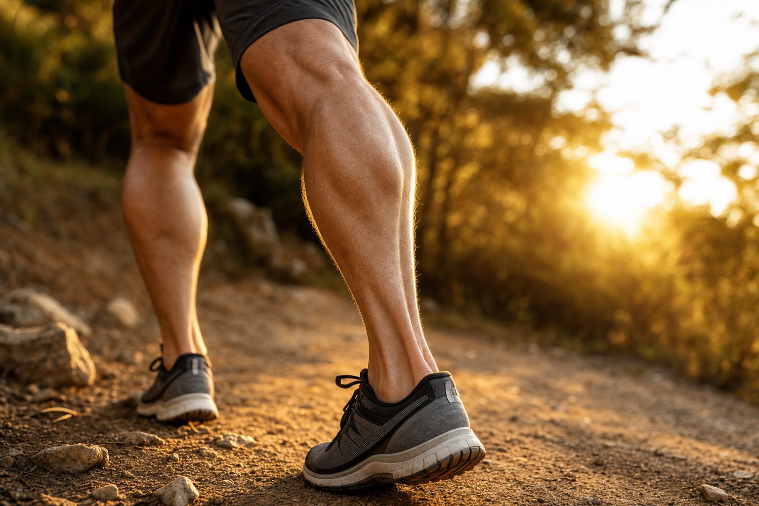
Tendon injuries are some of the most frustrating problems in sports and orthopedic medicine. A patient tears their Achilles — surgical repair is performed — and a year later, retear rates in some series run 20–30%. Another has chronic rotator cuff tendinopathy for 18 months; cortisone provides temporary relief, then the pain returns. Someone else has plantar fasciitis that has persisted for three years despite everything conservative medicine has to offer. The reason isn’t a failure of current clinical practice — it’s basic biology. Understanding what makes tendons structurally distinct from other tissues, and why this makes them inherently poor healers, is essential to understanding what regenerative medicine can and cannot do about it.
The Biology of Tendons: Why They Don’t Heal
Tendons are dense connective tissue structures that transmit force from muscle to bone — built from Type I collagen, tightly packed and parallel-aligned, providing the tensile strength to manage forces of 10–12x body weight during running and jumping. What makes tendons functionally brilliant makes them biologically fragile. There are four interlocking reasons why.
1. Poor Vascularization
Tendons have one of the lowest blood supplies of any connective tissue in the body. Critical regions — the rotator cuff insertion, the mid-portion of the Achilles, the proximal patellar tendon — receive blood supply only from peritendinous tissue, not directly from within the tendon itself. These avascular “watershed zones” are exactly where chronic tendinopathy and full-thickness tears most frequently occur. Without adequate blood supply, the body cannot efficiently deliver nutrients, oxygen, or healing cells to the injury site. This is the fundamental bottleneck in tendon repair biology.
2. Type I vs. Type III Collagen Dynamics
Healthy tendons are built from organized, mature Type I collagen — strong, parallel-aligned, and capable of bearing high loads. When injured, the initial repair response deposits Type III collagen — weaker, disorganized, and more elastic. Under normal biological conditions, Type III should gradually remodel to Type I over 12–18 months. In chronic tendinopathy, this remodeling is incomplete, arrested, or absent entirely. The structural consequence: tissue that looks healed on a clinical timeline but remains biomechanically inferior to the original.
3. Hypocellularity
Tendons have a low density of tenocytes — the resident cells responsible for collagen synthesis and matrix maintenance. When injury occurs, the limited local tenocyte population is insufficient to mount an adequate repair response on its own. External cell sources — from peritendinous tissue, circulating progenitor cells, or injected biologics — are needed to supplement native healing capacity. This is one of the core biological rationales for orthobiologic interventions: not replacing the tissue, but augmenting a repair environment that cannot self-supply adequate cellularity.
4. Tendinopathy Is Not Tendinitis
Acute tendon injury produces inflammation — tendinitis. Chronic tendon conditions — tendinopathy — are characterized by degeneration without active inflammation: disorganized collagen, mucoid ground substance, pathological neovascularization, and proliferating tenocytes attempting repair without achieving it. Treating tendinopathy with anti-inflammatory agents (cortisone, NSAIDs) is biologically counterproductive when chronic degeneration rather than active inflammation is the primary pathology. This is why cortisone provides short-term symptomatic relief — it suppresses the pain signal — but worsens long-term tendon integrity. The inflammation being suppressed is part of what remains of the healing response.
The Evidence for Regenerative Medicine by Tendon Location
The evidence base for PRP in tendinopathy is not uniform across anatomy. Understanding which tendons respond most reliably, what preparation variables matter, and where the evidence is genuinely mixed is essential for informed treatment decisions.
Rotator Cuff
The rotator cuff has the strongest evidence base for PRP of any tendon location. A 2025 systematic review and meta-analysis published in the Journal of Orthopaedic Case Reports (30 RCTs, approximately 2,500 participants) found PRP for rotator cuff tendinopathy “significantly reduced pain and improved function in the short term compared to placebo and corticosteroids.” A March 2026 Journal of Clinical Medicine study confirmed significant clinical improvement after PRP for supraspinatus tendinopathy. For surgical rotator cuff repair, the evidence is even more striking: a meta-analysis of 1,359 patients found PRP applied at the time of repair reduced retear rates from 23.6% to 16.5% (P=0.002). That is a meaningful reduction in a condition where retear is the primary driver of long-term failure.
Achilles Tendon
The evidence for isolated Achilles tendinopathy is more mixed — but more nuanced than a simple positive or negative verdict. A 2026 Journal of Clinical Medicine study found Achilles patients showed “the greatest improvement” after PRP within their cohort. Other meta-analyses found no significant improvement versus placebo in some series — a discrepancy that reflects the importance of leukocyte content, number of injections, and adjunct loading protocol rather than a fundamental failure of biology. The practical framework: eccentric loading is the foundation of Achilles tendinopathy management; PRP provides additive benefit. ESWT combined with PRP shows more consistent results than PRP alone for mid-substance Achilles tendinopathy.
Plantar Fascia
Plantar fasciitis has strong evidence supporting PRP over both corticosteroids and ESWT as a standalone intervention. A 2024 Foot and Ankle Surgery meta-analysis found PRP “significantly more effective than CSI, ESWT, and placebo in reducing VAS pain scores” — standardized mean difference versus corticosteroid of 1.08 (95% CI 0.05–2.11, p=0.04). A 2024 Cureus retrospective of 140 patients found PRP effective in 131 patients (93.57%), with age the only significant predictor of outcome. These are unusually strong numbers for a conservative intervention in a condition that routinely frustrates patients after years of standard care.
Patellar Tendon
The patellar tendon represents one of the more difficult tendinopathy targets. The 2026 Journal of Clinical Medicine study noted “reduced efficacy” for PRP at the patellar tendon compared to rotator cuff and Achilles findings. This aligns with clinical experience — the patellar tendon, with its high mechanical demands and proximity to the stress riser at the distal patellar pole, creates an environment where isolated PRP often underperforms. The most evidence-supported approach combines ESWT prior to PRP with eccentric decline-squat loading as the rehabilitation backbone. The combination matters here in ways that matter less for other tendons.
The Role of Leukocyte Content: LP-PRP vs. LR-PRP
One of the most clinically significant variables in PRP preparation — and one that most patients are never told about — is leukocyte content. Leukocyte-rich PRP (LR-PRP) contains white blood cells with concentrated inflammatory cytokines. Leukocyte-poor PRP (LP-PRP) is a more purified preparation with a lower inflammatory burden. For tendons specifically, LP-PRP appears to be the superior preparation — the inflammatory signal in LR-PRP may be counterproductive in chronically degenerated tissue where the pathology is degenerative, not inflammatory. Multiple studies and the 2026 JCM review suggest LP-PRP performs better for rotator cuff applications. This is a preparation detail that significantly affects clinical outcomes — and that many clinics offering generic “PRP injections” do not optimize or even acknowledge.
“Cortisone works for acute flares. For chronic tendinopathy, the long-term evidence is deeply unfavorable. PRP works with the biology of tendons in ways that cortisone never did.”
The Gap in Standard Care
The corticosteroid reflex for tendon pain is supported by decades of clinical habit. It is increasingly poorly supported by the evidence. Multiple studies and meta-analyses consistently document the same pattern for Achilles tendinopathy, plantar fasciitis, and rotator cuff tendinopathy:
- Corticosteroid injections provide better short-term pain relief than PRP at 4–6 weeks
- Corticosteroid injections produce inferior outcomes compared to PRP at 6–12 months
- Repeated corticosteroid injections are associated with tendon degeneration, collagen disruption, and increased long-term risk of rupture
The structural problem is that short-term outcomes dominate most follow-up schedules. Patients feel better at 6 weeks, get discharged, and return a year later with a worse tendon and often a worse prognosis. A comparison that tracks at 12 months consistently favors PRP and loading-based protocols over cortisone. The decision to choose cortisone is often a decision to optimize the 6-week visit at the expense of the 12-month outcome — and patients are rarely given that framing explicitly when the injection is offered.
This is not a fringe position. It reflects the current consensus in evidence-based sports medicine literature. The cortisone reflex persists not because the evidence supports it for chronic tendinopathy — it does not — but because it is fast, reimbursable, and produces short-term patient satisfaction scores that reinforce the behavior. A consultation at Pravida Health will give you a direct, biology-grounded conversation about where cortisone is appropriate and where it is not.
How We Use This at Pravida Health
Our approach to tendinopathy integrates four principles that distinguish a regenerative protocol from a generic “PRP injection” appointment:
- Accurate diagnosis with musculoskeletal ultrasound. Every tendon is assessed with ultrasound before treatment — tendon thickness, intratendinous hypoechogenicity, Doppler neovascularization, partial or full-thickness tears, calcification, and adjacent bursa pathology. A tendon with a 30% partial-thickness tear at the insertion is managed differently from diffuse mid-substance tendinopathy. Imaging before injection is not optional — it changes the clinical decision for every patient. Book a consultation to discuss your specific ultrasound findings.
- Leukocyte-matched PRP preparation. LP-PRP or LR-PRP based on specific clinical indication — preparation is matched to the pathology, not generic. For rotator cuff and most tendinopathies, LP-PRP is our standard. For wound healing or some intra-articular applications, LR-PRP may be more appropriate. The distinction matters clinically and we explain it to every patient.
- Combination with mechanical loading. Growth factors from PRP provide the biological signal for repair; eccentric loading provides the mechanical environment required for Type I collagen synthesis and fiber alignment. Both are required. PRP without a structured loading protocol produces inconsistent outcomes. We prescribe specific loading protocols — eccentric decline squats for patellar, eccentric heel drops for Achilles — as a mandatory component of every tendon treatment plan.
- ESWT integration when appropriate. Extracorporeal shockwave therapy (ESWT) prior to PRP is particularly valuable for chronic tendinopathy with significant fibrosis or calcific deposits. ESWT disrupts pathological calcification, stimulates mechanotransduction, and improves the local tissue environment — creating a more receptive substrate for the biological signal PRP delivers. Explore the full list of conditions we treat with this combined approach.
What You Can Do Today
- Stop cortisone after the second injection. One or two injections in an acute flare is defensible — short-term relief to allow rehabilitation to begin has legitimate clinical utility. Beyond that, the evidence strongly suggests you are trading structural integrity for short-term comfort. Discuss your cortisone history and whether a regenerative protocol is appropriate for your specific tendon and stage of pathology.
- Start eccentric loading. The evidence for eccentric exercise in Achilles and patellar tendinopathy is as strong as any pharmaceutical intervention. Eccentric decline squats for patellar tendinopathy; eccentric heel drops for Achilles — free, foundational, and undersold by most clinicians managing these conditions. They are not a consolation prize for patients who can’t access injection therapy. They are the mechanism of collagen remodeling.
- Use imaging before injection. PRP and ESWT should be guided by musculoskeletal ultrasound — both for diagnosis and accurate needle placement. An injection that misses the pathological zone of the tendon provides no regenerative benefit. Ultrasound guidance is not a premium add-on; it is how the procedure is done correctly.
- Allow adequate healing time. Tendons heal on a slow biological timeline — 3–6 months for meaningful biological change, 12–18 months for full Type III to Type I remodeling. Patients who assess outcomes at 4–6 weeks are measuring the cortisone-equivalent response, not the regenerative one. Commitment to the timeline is part of the protocol. See the conditions we treat and what a complete regenerative orthopedics plan looks like from diagnosis to return to activity.
Frequently Asked Questions
Why do tendons keep getting re-injured after treatment?
The healed tissue — Type III collagen scar — isn’t as strong or organized as original Type I collagen. Until remodeling produces mature, aligned Type I collagen, the tendon remains biomechanically vulnerable. Adequate rehabilitation time (minimum 6 months for full remodeling), gradual load progression, and optimizing the healing environment with orthobiologics reduces but does not eliminate this risk. This is why return-to-sport timelines after tendon injury are longer than patients typically expect — and why cutting them short is the most common cause of re-injury.
Is PRP better than surgery for a rotator cuff tear?
It depends on tear size and chronicity. For partial-thickness tears, PRP — often combined with ESWT — is a legitimate non-surgical option with strong supporting evidence. For full-thickness tears greater than 1.5 cm, surgical repair generally produces better anatomical outcomes. PRP at the time of surgery reduces retear rates significantly — the meta-analysis (n=1,359) documenting a reduction from 23.6% to 16.5% is compelling. Surgery and PRP are not competing options in the right context. They are complementary: surgery restores anatomy; PRP optimizes the biological healing environment.
How long does PRP take to work for tendinopathy?
Early response — pain reduction and reduced swelling — often begins at 2–4 weeks. Meaningful functional improvement is typically seen at 6–12 weeks. Maximum biological effect — collagen remodeling and structural improvement — peaks at 3–6 months. Patients who assess outcomes at 4–6 weeks are measuring the cortisone-equivalent response, not the regenerative one. The biological timeline for tendon healing is slow by design — managing expectations around that timeline is part of responsible care.
Can I get PRP for both Achilles tendinopathy and knee OA at the same visit?
Yes. Drawing sufficient blood for multiple PRP preparations is feasible in a single session. We regularly treat adjacent pathologies — or pathologies at multiple sites — in the same visit when indicated clinically. It is efficient, cost-effective, and biologically sound. Each preparation is still matched to its specific clinical indication.
What tendon and ligament conditions does Pravida Health treat?
Our regenerative orthopedics program addresses chronic Achilles tendinopathy, plantar fasciitis, patellar tendinopathy, lateral and medial epicondylosis (tennis and golfer’s elbow), rotator cuff tendinopathy and partial tears, gluteal tendinopathy, and ligamentous injuries amenable to PRP. Every case is evaluated with musculoskeletal ultrasound before any injection is performed. See the full list of conditions we treat for more detail on how each is approached.
Dealing with a chronic tendon problem that hasn’t responded to cortisone, rest, and PT?
A consultation at Pravida Health begins with musculoskeletal ultrasound, a biology-first assessment of your specific tendon pathology, and an honest conversation about whether a regenerative protocol is appropriate for your stage of disease. No generic injections. No shortcuts on the timeline. A physician-led plan that works with tendon biology rather than against it.
Book a Consultation Explore Conditions We Treat