
Series introduction: the spine and joint as a symphony
Almost every patient who comes to me for a regenerative procedure has already had “a shot” somewhere else — sometimes several. Usually the same shot, in roughly the same place, delivered under roughly the same imaging. When it works for a while and stops working, the assumption is that the injection was wrong, or the medicine was wrong, or the diagnosis was wrong. That is occasionally true. Far more often, what was wrong is that the joint or spine segment was being treated as if it were one part, when it is actually an orchestra.
Every functional unit in the musculoskeletal system — a knee, a shoulder, a lumbar segment — is a coordinated ensemble of structures that only produces smooth, painless motion when each section plays in tune with the others. There is bone. There is cartilage. There are ligaments that stabilize. There are muscles that generate force. There is fascia that transmits it. There are nerves that report on all of it in real time. When one section falls out of tune — a stretched ligament, an atrophied stabilizer, a sensitized nerve root, a scarred sheet of fascia — the whole ensemble sounds off.
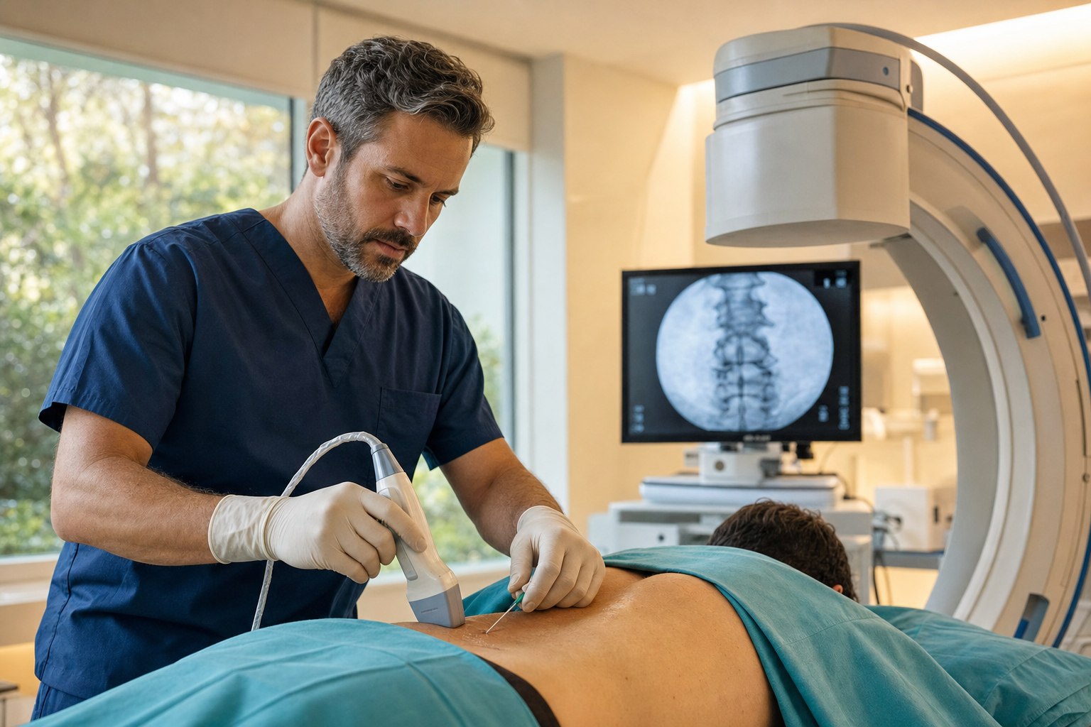
A world-class regenerative procedure, in my hands, is the conductor’s approach to that ensemble. It is not one injection. It is the deliberate treatment of every structure that is out of tune, using the imaging tool that shows that structure best — fluoroscopy for what lives deep and against bone, ultrasound for what lives in soft tissue where the needle needs to be seen moving in real time. This article is the first in a series that walks through each region I treat this way. We start where I get the most calls: the lumbar spine.
Why two imaging tools, and not just one
The single most common question I hear about our approach is a version of “why do you use both fluoroscopy and ultrasound?” The short answer is that they see different things.
Fluoroscopy is real-time X-ray. It excels at showing bone and, with a small amount of contrast dye, the flow of medication through spaces that live deep against bone — the neural foramen where a nerve root exits the spine, the inside of a lumbar disc, the epidural space. Fluoroscopy is how we prove, on a live image, that the needle tip is exactly where we want it before we deliver anything, and how we watch contrast fill the target space along the correct anatomic pathway. For any procedure that reaches structures behind and between the vertebral bones, fluoroscopy is the standard of care.
Ultrasound is real-time sound imaging. It cannot see through bone, but it excels at exactly what fluoroscopy cannot show — soft tissue. Muscle fibers, ligaments, fascial layers, nerves in the periphery, blood vessels. On ultrasound I can watch a needle advance millimeter by millimeter through a specific fascicle of the multifidus muscle, or into the precise interspinous space between two spinous processes, and I can see the biologic spread through the tissue as it is delivered. For soft-tissue targets, ultrasound gives me a level of precision and safety that no other tool matches.
A physician who owns only one of these tools has to force every problem into what that tool can see. A physician who owns both can choose the right tool for each structure. That is the practical, unglamorous reason a comprehensive lumbar procedure at Pravida usually involves both imaging systems in the same room.
The five sections of the lumbar orchestra
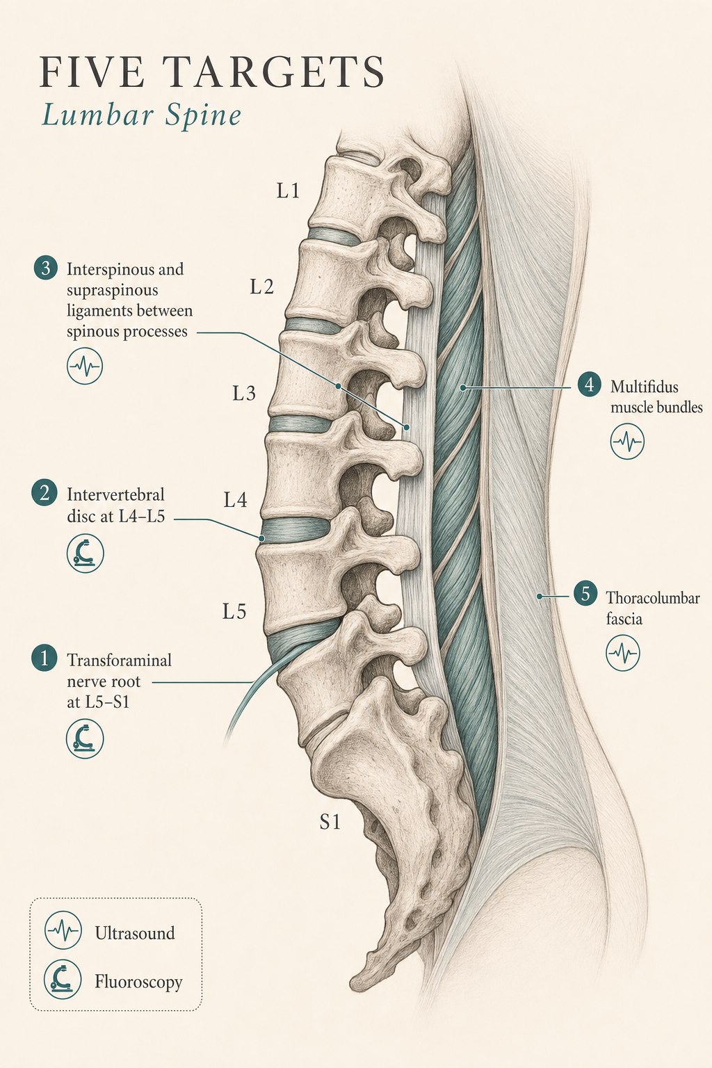
What follows is a section-by-section walk through the five structures I most commonly treat in a comprehensive lumbar procedure. For each one I describe what the structure actually does when it is playing in tune, how it manifests as pain when it falls out of tune, and how it is targeted during the procedure — including which imaging tool is right for that particular section.
1. The lumbar nerve root — the electrician of the leg
What it does: Each lumbar nerve root exits the spine through a small bony tunnel called the neural foramen. It carries motor signals to a specific set of leg muscles and sensory information back from a specific patch of skin. The L5 root, for example, drives the muscle that lifts your foot at the ankle and reports sensation from the top of the foot. When it is healthy, you do not notice it at all — the electrician is invisible when the lights are on.
How it manifests as pain: When a disc herniates and presses on a nerve root, or when a foramen narrows with age and pinches it, the nerve becomes chemically and mechanically inflamed. The pain is unmistakable to anyone who has had it — a sharp, electric, unmistakably nerve-quality pain that travels from the low back into the buttock and down the leg, often below the knee. This is what most people mean when they say “sciatica.” It may be accompanied by numbness in the same distribution or, in more serious cases, weakness in the specific muscles that nerve root drives.
How we target it: With a fluoroscopically guided transforaminal epidural injection. Under live X-ray, we advance a thin needle to the outer edge of the neural foramen where the nerve root exits, confirm position with a small amount of contrast dye, and deliver medication directly to the sleeve of the nerve root and the epidural space around it. This is the most precise approach we have to a specifically inflamed nerve root, with Level I evidence for radicular pain from disc herniation and satisfactory long-term outcomes in the range of eighty percent in appropriately selected patients (Kim et al., 2025). In regenerative practice, we can pair or replace the traditional corticosteroid with autologous biologics where indicated. The tool is fluoroscopy specifically because the neural foramen lives deep against bone, in a space where ultrasound simply cannot see with the fidelity required.
2. The intervertebral disc — the shock absorber
What it does: Between each pair of lumbar vertebrae sits a disc — a tough outer ring of concentric fibers (the annulus fibrosus) surrounding a hydrated inner core (the nucleus pulposus). Together they act as a hydraulic shock absorber and a pivot point, allowing the spine to bend, compress, and rotate without transmitting the full load directly onto the bony vertebrae above and below.
How it manifests as pain: Discogenic pain is deep, midline, dull, and axially loaded. It is worse with sitting (the position that puts the highest compressive load through the disc), better with walking, and often described as a “band of ache” across the low back rather than a specific point. As a disc degenerates, tiny fissures develop in the annulus that allow small nerve fibers to grow inward — the disc itself becomes innervated in a way it should not be, and it starts to report as a pain generator. This can happen with or without a frank disc herniation.
How we target it: With a fluoroscopically guided intradiscal injection. Under live X-ray we advance a needle through a specific anatomic corridor into the center of the affected disc, confirm intradiscal position with contrast, and deliver a regenerative biologic directly into the nucleus. Intradiscal orthobiologic therapy is one of the most technically demanding procedures in interventional pain medicine and one of the most rewarding when done well — it is the closest we have to disease-modifying therapy for the disc itself, rather than symptom management around it. The tool is fluoroscopy for the same reason as the nerve root: the disc lives at the center of the spine, deep against bone, in a space where only live X-ray gives us the safety margin we need.
3. The interspinous, supraspinous, and iliolumbar ligaments — the tension cables
What they do: A ligament’s job is to control the endpoint of a joint’s motion — to allow full motion up to a normal limit, then stop it cleanly. The lumbar spine has a small set of ligaments that matter enormously for stability. The interspinous and supraspinous ligaments run between and along the bony spinous processes you can feel down the midline of your back — they resist excessive forward bending. The iliolumbar ligament runs from the transverse process of L5 (and sometimes L4) out to the top of the iliac crest — it is the tension cable that anchors the base of the lumbar spine to the pelvis (Physiopedia review).
How they manifest as pain: When these ligaments are stretched, thickened (hypertrophied), or torn from a specific mechanism — a fall, a heavy asymmetric lift, chronic instability of a lumbar segment — they become pain generators in a very characteristic pattern. Iliolumbar ligament pain is one of the most under-diagnosed causes of chronic low back pain: unilateral, deep, right over the posterior iliac crest, without radiation into the leg, worsened by prolonged sitting or standing and by side-bending toward the opposite side. Interspinous ligament pain is midline, provoked by end-range forward bending, and tender to direct palpation over the spinous processes. In both cases patients have often been told they have “a disc” when they in fact have a ligament that has never been treated.
How we target them: With ultrasound-guided injections. Ligaments are soft-tissue structures that live under the skin at very shallow depth — the interspinous ligament is sometimes only a centimeter below the surface, the iliolumbar ligament runs at an oblique angle that only ultrasound can reliably visualize. On ultrasound I can identify the specific fascicle of the ligament that is hypertrophic or scarred, watch the needle enter it, and deliver a biologic in a way that would be blind on fluoroscopy. This is where the two-tool approach pays off: fluoroscopy would give me a picture of the bony landmarks, but only ultrasound can show me the ligament itself.
4. The multifidus — the deep segmental stabilizer
What it does: The multifidus is a small, deep muscle that runs in short segments along the back of the spine, from each vertebra to the ones one or two levels below. Unlike the big superficial back muscles that move the trunk, the multifidus stabilizes each individual segment so that the vertebrae above and below cannot move on each other without permission. If the spine is a chain, the multifidus is the small clip that holds each link in place. It fires in a feed-forward pattern — a hundred milliseconds before you move your arm or your leg — to preemptively stiffen the segment against the load that is about to arrive.
How it manifests as pain: When a lumbar segment becomes painful for any reason — a disc, a facet joint, a nerve root — the multifidus at that segment goes through a well-described process of reflex inhibition. The nervous system shuts down its firing to protect the painful segment from further loading, and within weeks the muscle begins to shrink. Over months it becomes replaced by fat — a process visible on MRI as fatty infiltration. This has been documented in a series of imaging studies showing that a majority of patients with chronic low back pain have measurable multifidus atrophy at the involved segment, and the classic paper by Hides et al. showed the atrophy can develop within days to weeks of a single episode of back pain. Once the stabilizer is out of the ensemble, every other structure at that segment is overloaded, and the pain becomes self-perpetuating.
How we target it: With ultrasound-guided injection directly into the multifidus fascicles at the involved segments. Under ultrasound we can identify the specific fatty, atrophic-appearing muscle bundle, watch the needle enter it, and deliver a biologic that supports the local environment while a targeted rehabilitation program — on our OxeFit platform and with our physical therapy partners — reactivates the muscle’s firing. The point of injecting the multifidus is not that a shot rebuilds a muscle. It is that a shot can help unlock the reflex inhibition and quiet the local inflammation enough that the muscle can be trained again. Every serious regenerative spine practice I respect is doing some version of this now, because ignoring the multifidus is one of the reasons single-target injections stop working.
5. The thoracolumbar fascia — the pain-sensitive canvas
What it does: The thoracolumbar fascia is a broad, layered sheet of connective tissue that spans the back from below the ribs to the pelvis. It is where several muscle groups — latissimus dorsi, gluteus maximus, the deep spinal muscles — attach and transmit force. Think of it as the canvas onto which the low-back and hip muscles paint. When it is healthy, it glides smoothly between its layers and transmits force from the hip to the trunk (and back) with almost no loss.
How it manifests as pain: Experimental work by Siegfried Mense and others has established that the thoracolumbar fascia is densely innervated with nociceptive nerve endings, containing calcitonin gene-related peptide and substance P-positive free nerve endings similar to those in a peripheral nerve. In healthy volunteers, direct injection of a small volume of hypertonic saline into the thoracolumbar fascia produces some of the most intense and long-lasting experimental low-back pain of any tissue tested — more than muscle, more than ligament (Schilder et al.). Clinically, thoracolumbar fascia pain is a broad, diffuse, myofascial ache across the lower back, worse with static postures, often described by patients as “my back is tight” when it is really the fascia that is sensitized. When the fascia becomes scarred or stuck between its layers — from prior injury, prior surgery, or chronic overload — the canvas can no longer glide, and it becomes a chronic pain generator.
How we target it: With ultrasound-guided injection into the specific fascial layer that shows scarring or restricted glide on dynamic imaging. Ultrasound is the only imaging tool that can visualize the multiple thin layers of the thoracolumbar fascia in real time and, importantly, can watch them glide (or fail to glide) as the patient breathes and shifts. On fluoroscopy a fascial injection is a leap of faith. On ultrasound it is a precisely placed hydrodissection that separates a stuck fascial plane and delivers a biologic to the tissue that is actually the pain generator. This is the section of the lumbar orchestra that is most consistently missed by traditional single-target injection practices — and it is often the one that finally quiets the chronic myofascial background pain patients have lived with for years.
How the conductor puts the ensemble together
A comprehensive lumbar regenerative procedure at Pravida does not treat all five of these structures on every patient. The point of the physical exam, the imaging review, and the diagnostic history is to determine which sections of the ensemble are actually out of tune for you. Some patients will have a clear single-structure problem — a discretely herniated disc pressing on an L5 root — and a single well-placed transforaminal epidural is exactly the right answer. Other patients will have three or four of these structures contributing simultaneously, and treating just one is why the last three injections wore off in six weeks.
What a world-class procedure looks like on the day it happens: a physical exam and imaging review that identifies the specific structures in play, a written plan for which structures will be treated, an operating room set up with both a C-arm fluoroscope and a high-resolution ultrasound machine, and a sequence of small, precise injections — each one confirmed on real-time imaging before medication is delivered — that treats each identified section of the ensemble with the tool that shows it best. The patient is usually mildly sedated but comfortable and responsive. The whole procedure typically takes forty-five to seventy-five minutes depending on how many structures are involved, and most patients walk out of the clinic the same afternoon.
The choice of biologic — platelet-rich plasma, bone marrow concentrate, ev-enhanced preparations, or in some cases a supplemental non-regenerative agent for a specific structure — is guided by the tissue being treated and by the patient’s overall regenerative plan. The CartiNova program is our organized framework for making those choices in a way that is transparent, evidence-informed, and personalized to the individual patient.
What most patients get wrong before they see us
- They assume “the shot” is one thing. A single well-placed injection into one structure is often the right first step, but when it wears off, the answer is usually not to repeat the same injection — it is to widen the diagnostic lens to include the other sections of the ensemble.
- They confuse imaging findings with pain generators. A herniated disc on an MRI is not, by itself, a pain diagnosis. Twenty percent of asymptomatic forty-year-olds have a disc herniation. The question is not what is on the MRI — it is which structure is producing your specific pain pattern today.
- They ignore the multifidus and the fascia. These are the two structures most consistently missed by conventional injection practices. If your prior injections have targeted only the nerve root or only the disc and stopped working, ask about these two.
- They expect regeneration without rehabilitation. A biologic injection into an inhibited multifidus does not rebuild the muscle. It creates a window in which targeted rehabilitation can rebuild the muscle. The injection and the rehab are one intervention, not two.
What this series will cover next
This is the first article in a series on how a master regenerative procedure actually works, region by region. Future articles in the series will apply the same conductor’s approach to the cervical spine, the shoulder, the hip, the knee, and the elbow, wrist, and foot — each with its own five-to-eight-structure ensemble and its own map of when fluoroscopy is the right tool and when ultrasound is. The framework is always the same: identify every section of the orchestra that is out of tune, treat each one with the imaging modality that shows it best, and build a regenerative plan that treats the joint or spine segment as a functional unit rather than a single part.
The honest limits, and where a good clinician still matters
- The five-structure approach is designed for patients with a chronic or subacute mechanical lumbar pain pattern that has not fully resolved with conservative care. It is not a first-line treatment for acute new-onset radicular pain (a trial of conservative care and, when indicated, a targeted epidural comes first), for red-flag presentations (see below), or for suspected surgical emergencies.
- Not every patient needs every structure treated. Some patients need one structure, and adding more injections to a patient who has a single-structure problem is not more thorough — it is more expensive and adds unnecessary risk. The physical exam and imaging review are what decide.
- Regenerative outcomes depend on the underlying tissue, the biologic used, the technical precision of delivery, and the rehabilitation that follows. A biologic delivered blindly, or into the wrong structure, is not regenerative — it is just an injection. Precision is not optional in this work.
- If you have new leg weakness, saddle numbness, loss of bladder or bowel control, unexplained fever or weight loss, or back pain following significant trauma, please contact your physician or an emergency department today. Those are not situations for an elective regenerative procedure.
Ready to have your specific lumbar pattern mapped?
If you are an Atlanta-area patient with chronic low back pain that has not fully responded to prior injections or conservative care — or you want a second opinion on which of these five structures is actually driving your pain — we would rather see you in person. A consultation at Pravida Health includes a physical exam, an imaging review, and, where appropriate, a comprehensive regenerative plan tailored to the specific sections of your lumbar orchestra that are out of tune.
Book a consultationKey sources referenced in this article
- Kim H, et al. Long-term outcomes of transforaminal epidural steroid injection for lumbar radicular pain: a systematic review. 2025. PMC12422987. Reports satisfactory outcomes at two years in approximately eighty percent of appropriately selected patients.
- Systematic review, transforaminal epidural steroid injection for lumbar radicular pain from disc herniation. 2025. PubMed 40908263. Level I evidence supporting transforaminal epidural for radicular pain of disc origin.
- Multifidus atrophy and fatty infiltration in chronic low back pain: imaging and clinical review. PMC12862208. Discusses the prevalence of multifidus atrophy in chronic low back pain and the biomechanical consequences.
- Hides JA, Stokes MJ, Saide M, Jull GA, Cooper DH. Evidence of lumbar multifidus muscle wasting ipsilateral to symptoms in patients with acute/subacute low back pain. Spine. 1994;19(2):165–172. The original paper documenting segmental multifidus atrophy after a single episode of low back pain.
- Mense S. Innervation of the thoracolumbar fascia. Eur J Transl Myol. 2019. PMC6767935. Anatomical review demonstrating dense nociceptive innervation of the thoracolumbar fascia with CGRP- and substance P-positive free nerve endings.
- Schilder A, et al. Sensory findings after stimulation of the thoracolumbar fascia with hypertonic saline suggest its contribution to low back pain. Pain. 2014. PMC5444000. Experimental evidence that the thoracolumbar fascia is one of the most pain-sensitive structures in the low back.
- Physiopedia. Iliolumbar ligament: anatomy, function, and clinical presentation. physio-pedia.com/Iliolumbar_ligament. Reference summary of the iliolumbar ligament’s role in lumbopelvic stability and its characteristic pain pattern.