
The problem that has never really been solved
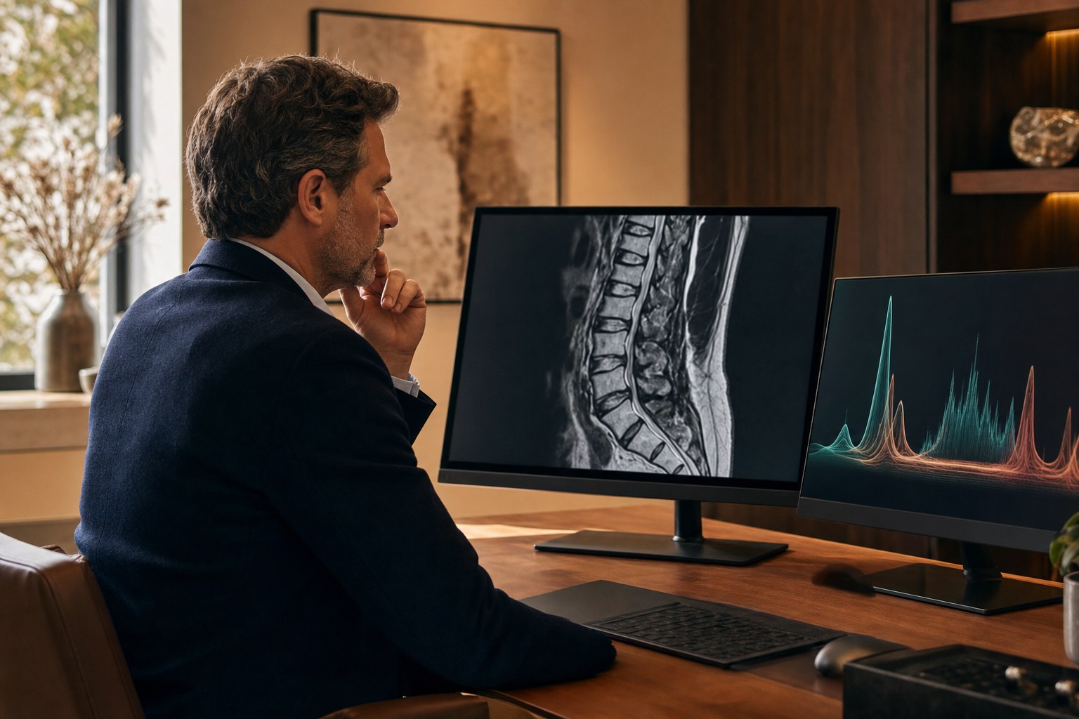
A patient comes to me with four years of axial low back pain. It is worse with sitting, worse with flexion, better lying down, and it has not responded to a good course of physical therapy. I order a lumbar MRI. The MRI comes back and it shows disc desiccation and loss of height at L4–L5 and L5–S1, a small annular fissure somewhere in there, maybe a Modic change, and some facet arthropathy for good measure.
Now I have a problem that is much older than I am. Which of those discs, if any, is generating the pain?
This is not a rhetorical question, and it is not a small one. If I am considering an intradiscal regenerative procedure — CartiNova, intradiscal platelet-rich plasma, or bone marrow aspirate concentrate — I am about to put a needle into a specific disc and inject something into it. The value of that procedure depends almost entirely on whether I chose the right disc. Structural findings on MRI are notoriously poor at telling me which one. Degenerative changes are extremely common in people with no back pain at all, and severity of imaging findings correlates weakly with symptoms.
So for decades the answer has been provocative discography: insert a needle into each candidate disc, pressurize it with contrast, and see whether it reproduces the patient’s pain. It is a functional test rather than a structural one, and conceptually it is the right idea. Practically, it is invasive, it is uncomfortable, it depends heavily on subjective patient report during the procedure, it requires needles in multiple discs including presumed-normal control levels, and there is a long-running and genuinely unresolved debate about whether puncturing a disc accelerates its degeneration. It has fallen out of favor in a lot of practices, mine included, and it is frequently not covered by insurance.
That has left a real gap. And it is the gap that magnetic resonance spectroscopy of the disc is trying to fill.
What MR spectroscopy of a disc actually measures
Most of us learned spectroscopy in the context of the brain — distinguishing a tumor from radiation necrosis, that sort of thing. The physics is the same here, applied to a much less cooperative target. Rather than producing an anatomic image, a spectroscopy sequence produces a chemical spectrum from a defined volume of tissue, showing the relative concentrations of specific metabolites.
Applied to a lumbar disc, the technology of interest is Nociscan, developed by Aclarion. It is a cloud-based software service rather than a piece of hardware: the imaging site acquires an MR spectroscopy sequence during an otherwise routine lumbar MRI, uploads the raw spectral data, and proprietary signal processing and algorithms return a report. There is no separate scanner to buy, which matters a great deal for how quickly a radiology group can adopt it.
The biology it is built on is straightforward, and I find it genuinely compelling. Two families of signal are quantified. The first is structure — proteoglycan and collagen, the load-bearing matrix of a healthy disc. The second is acidity and degradation — lactate, alanine, and propionate, the metabolic by-products that accumulate in a disc that has shifted toward anaerobic metabolism and inflammatory breakdown. In the pivotal work, ratios of acidity to structure were used to calculate what the authors term the pain potential of each disc, which the algorithm reports as NOCI-positive or NOCI-negative (Gornet et al., European Spine Journal 2019).
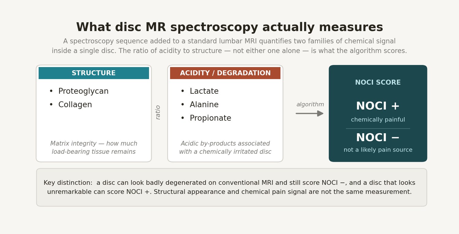
That last point is the entire clinical premise, and it is worth sitting with. Conventional MRI tells me what a disc looks like. This is attempting to tell me what a disc is doing chemically. Those are different questions, and the second one is much closer to the question I actually need answered before I plan a procedure.
The accuracy data, and where it comes from
The foundational study is Gornet and colleagues in European Spine Journal in 2019: 139 patients and 623 discs, with MR spectroscopy results compared against provocative discography as the reference standard. Across all discs, accuracy was 85%, sensitivity 82%, and specificity 88%. When the analysis was restricted to non-herniated discs — which is the population where the diagnostic question is hardest and most clinically relevant — those figures rose to 93% accuracy, 91% sensitivity, and 93% specificity (PubMed 30610465).
A later prospective cohort published in the International Journal of Spine Surgery in 2024 reported 87% accuracy, 100% sensitivity, and 80% specificity. That study, however, comprised just 14 patients and 44 discs, of which 4 discs were excluded for inadequate signal quality, and it was sponsored by Aclarion (Gornet et al., Int J Spine Surg 2024).
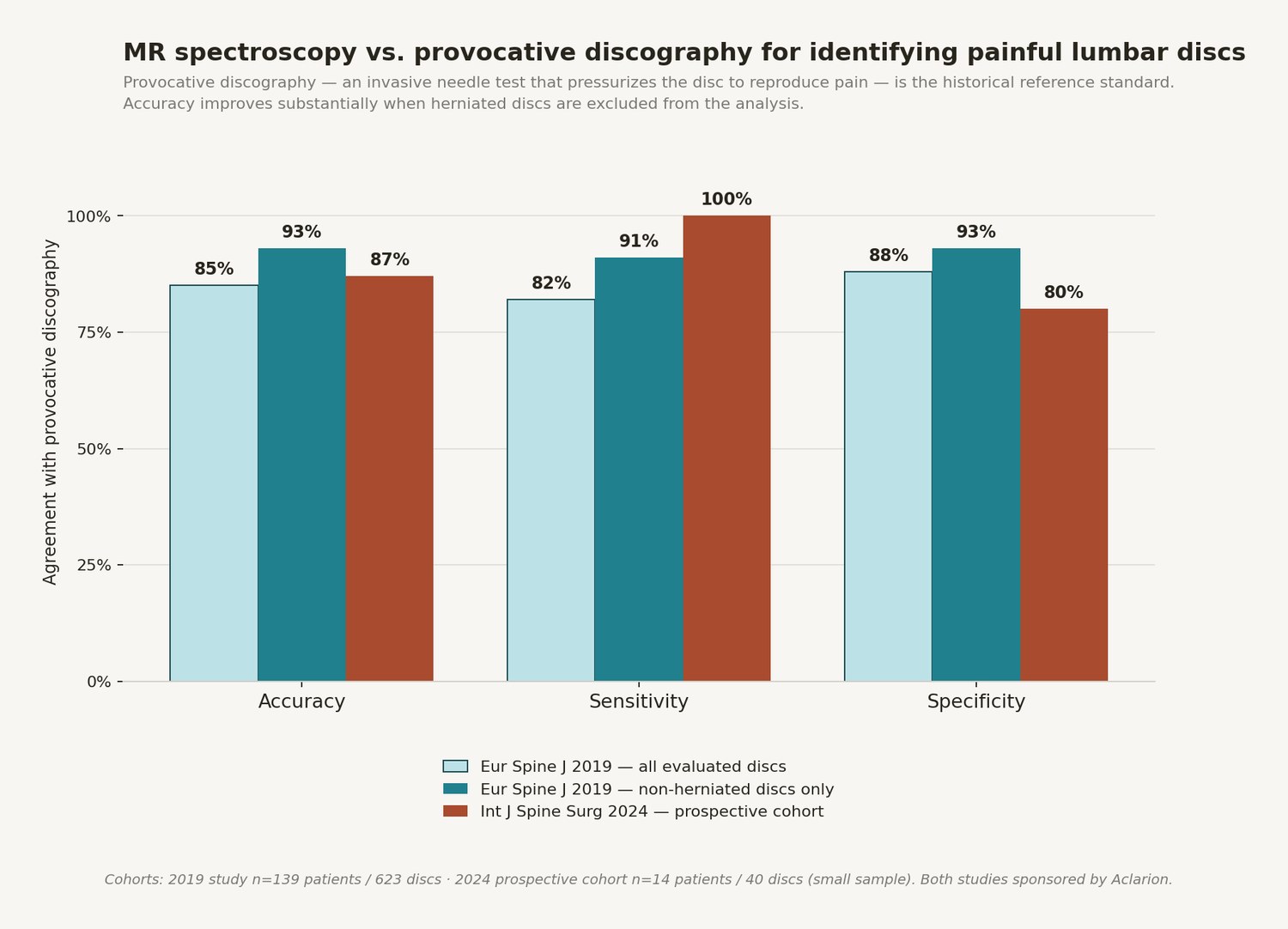
I want to flag a methodological point that is easy to skate past. These figures describe agreement with provocative discography, and discography is an imperfect reference standard with its own false-positive rate. A test can only look so good against a flawed comparator, and it can also be penalized for being right when the comparator was wrong. Agreement with discography is meaningful and it is the honest place to start, but it is not the same as being correct. Which is exactly why the outcome data matters more than the accuracy data.
A clarification about the “European” data
I have heard this technology discussed in terms of impressive European surgical data, and I had the same impression myself before I went through the primary literature. The distinction turns out to be worth making precisely.
The pivotal Gornet studies were published in European Spine Journal, which is the official journal of the EuroSpine society and a genuinely rigorous venue. The studies themselves, however, were conducted at United States sites — the Orthopedic Center of St. Louis, Scripps Clinic, and the University of California San Francisco. So the data is European by publication, not by geography.
That said, real European clinical activity does exist, and it is not trivial. The London Clinic has adopted Nociscan and has run roughly a hundred scans. Campus Bio-Medico University in Rome is running the ACTIVE trial, and Haukeland University Hospital in Bergen, Norway is running the LIFEHAB trial (Aclarion evidence page). Those are meaningful European academic engagements. But the numbers that made the strongest impression — the accuracy figures and the surgical concordance figures — came out of American operating rooms.
The result that actually changed my thinking
Diagnostic accuracy is a necessary claim but not a sufficient one. A test earns its place in a treatment algorithm only if using it leads to better outcomes. On that question, there is data, and it is the most interesting part of this story.
In the same 2019 cohort, the authors looked at surgical outcomes as a function of whether the operation matched what the spectroscopy had flagged. When all MRS-positive discs were treated, the surgical success rate was 97%. When the level that was actually treated had scored MRS-negative, success dropped to 57%. And when an adjacent level that had scored MRS-positive was left untreated, success was 54% (PubMed 30610465).
A follow-up study in European Spine Journal in 2023 examined this prospectively in 78 patients undergoing lumbar fusion or total disc replacement for discogenic pain, defining success as at least a 15-point improvement in Oswestry Disability Index. Patients whose surgery was concordant with the spectroscopy findings did better at every timepoint: 88% versus 62% at six months, 91% versus 56% at twelve months, and 85% versus 63% at twenty-four months (Gornet et al., Eur Spine J 2023).
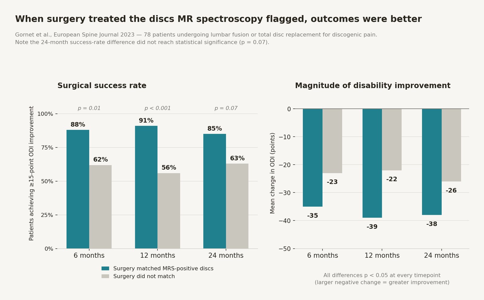
I want to be careful here rather than enthusiastic. That twenty-four-month figure did not reach statistical significance, at p = 0.07. A lot of promotional material about this technology quotes the twenty-four-month numbers without that caveat, and I think that is a mistake. The magnitude-of-improvement comparison held up better — mean ODI change was 38 points versus 26 points at twenty-four months, and the ODI change differences were significant at every timepoint — but the binary success-rate difference at two years is best described as a trend in a study of 78 patients.
It is also worth naming the inherent limitation of this study design. This is concordance analysis, not randomization. Surgeons chose their levels using their own judgment, and the spectroscopy was compared against those choices after the fact. It is entirely possible that the cases where surgical judgment and spectroscopy agreed were simply the clearer, more straightforward cases, and that they would have done better regardless. That is not a reason to dismiss the finding. It is a reason to want a randomized trial, which is exactly what is now underway.
What a randomized trial would settle
The CLARITY trial is a prospective, randomized, multicenter study enrolling up to 300 patients randomized one-to-one to surgical planning with or without spectroscopy data. The primary endpoint is change in back pain on a 100mm visual analog scale at twelve months. The principal investigator is Dr. Nicholas Theodore at the University of Arizona College of Medicine in Phoenix, and the trial is fully funded (NCT06661850).
This is the study that will determine whether the technology becomes standard of care or a footnote. Randomization removes the selection-bias problem entirely: if the group whose surgeons had the spectroscopy data does better, that difference is attributable to the information. I am watching it closely, and I would encourage anyone considering this technology to watch it too rather than treating the concordance data as settled.
The cost argument, which is stronger than I expected
A decision-analytic model led by Leslie Wilson at UCSF compared discography-based and spectroscopy-based diagnostic pathways over a twelve-month horizon in 2022 US dollars. Total cost per patient was $59,711 for the discography pathway versus $57,998 for the spectroscopy pathway — a saving of $1,712 per patient. The diagnostic test itself was modeled at $1,950 for discography versus $1,450 for spectroscopy, and the modeled surgical success rate was 78% with spectroscopy versus 68% with discography. Spectroscopy was the dominant strategy: less expensive and more effective (Wilson et al., cost-effectiveness analysis).
Modeling studies are only as good as their input assumptions, and the surgical success rates fed into this model came from the observational data discussed above. But the logic is intuitive even without the arithmetic: the expensive thing in spine care is not the diagnostic test, it is the operation that does not work. Anything that reduces the rate of unsuccessful procedures pays for itself quickly.
Why I care about this for regenerative procedures — and the limit I have to be honest about
Here is where my interest becomes specific, and where I need to be most careful.
The case for level selection is arguably stronger in regenerative medicine than in fusion surgery. When I inject a disc with CartiNova, intradiscal PRP, or BMAC, I am delivering a biologic to one specific structure. There is no adjacent-level stabilization, no incidental decompression, no mechanical effect on neighboring segments that might help by accident. If I inject the wrong disc, I have delivered a treatment to a tissue that was not the problem, and the patient has undergone a procedure for nothing. The intervention is more targeted, which means the target selection matters more.
That is a reasonable argument. It is also, at present, entirely a mechanistic argument.
The most important limitation in this article
- All published outcome data is surgical. Every outcome study on this technology to date has been conducted in patients undergoing lumbar fusion or total disc replacement.
- There is no published evidence that MR spectroscopy improves outcomes for CartiNova, intradiscal PRP, intradiscal BMAC, or any other regenerative disc procedure.
- Mechanistically reasonable is not the same as empirically demonstrated. The logic that better level selection should improve regenerative outcomes is sound, and it is untested.
- The direction of effect is not even guaranteed. It is conceivable that a chemically acidic, highly degraded disc is a worse environment for injected cells or platelets to survive in, and that a NOCI-positive disc predicts pain but not treatment response. Nobody has published data either way.
I raise that fourth point because it is the one I keep returning to. The biology that makes a disc chemically painful — low pH, lactate accumulation, matrix breakdown — is also biology that is hostile to cell survival. It is entirely possible that spectroscopy identifies the disc that hurts and simultaneously identifies the disc least likely to respond to a biologic. That would be an uncomfortable finding, and it would also be extremely useful to know. Until somebody studies it, anyone telling you that spectroscopy improves regenerative disc outcomes is extrapolating, including me.
For context on where the underlying regenerative evidence stands: intradiscal biologics remain an area of active investigation with evolving guidance, and systematic review of intradiscal bone marrow aspirate concentrate has covered on the order of sixteen studies and six hundred participants with encouraging but heterogeneous results (systematic review of intradiscal BMAC; intradiscal biologics guidance). We are layering an investigational diagnostic on top of an investigational therapeutic. That is not a reason not to do it thoughtfully, but it is a reason to be extremely clear with patients about what is known.
How I would actually use it
With all of that said, here is the decision framework I would apply. I want to emphasize that this is a decision map reflecting how the data is used in spine surgery, adapted to my setting — not a validated protocol.
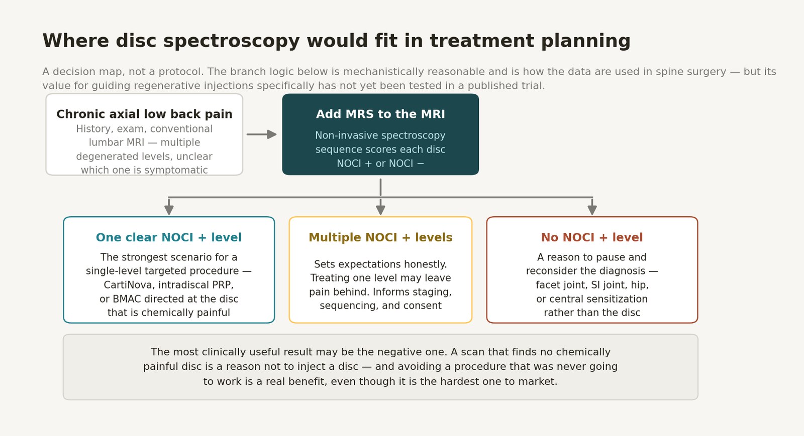
One clear NOCI-positive level. This is the scenario that most supports a single-level targeted procedure. I have an anatomic finding, a clinical picture, and a chemical signal that all point at the same segment. That is the strongest case I can build with the tools available.
Multiple NOCI-positive levels. This is less satisfying but arguably more valuable, because it sets expectations honestly. If two discs are chemically painful and I treat one, the patient may improve partially and be disappointed. Knowing that in advance changes the consent conversation, the staging plan, and what I tell them to expect. Patients tolerate partial results far better when they were told to expect them.
No NOCI-positive level. This is, I suspect, the most clinically useful result of all, and it is the one nobody markets. If a patient with chronic axial low back pain has no chemically painful disc, that is a strong signal to stop and reconsider the diagnosis before putting a needle anywhere near a disc. The pain may be facetogenic. It may be sacroiliac. It may be referred from the hip. It may be a central sensitization picture that no injection is going to solve. Avoiding a procedure that was never going to work is a real clinical benefit — it is just the hardest kind of benefit to build a business around.
If you want to talk through which of these scenarios your own imaging and symptoms fit, that is a reasonable thing to bring to a consultation.
Availability, and why Atlanta is a gap
Nociscan is currently available at a scattered set of sites, concentrated in a few metros. In New York, that includes multiple Lenox Hill Radiology locations and Weill Cornell’s Och Spine program. In the Los Angeles area, it includes the Institute of Neuro Innovation in Santa Monica, Lanman Spinal Neurosurgery in Beverly Hills, Medical Imaging Center of Southern California, Source Healthcare, and USC. The Dallas-area presence is in Plano, at Texas Back Institute and Texas Health Center for Diagnostics and Surgery. Additional academic sites include Johns Hopkins, Northwestern, the University of Miami, AdventHealth Porter in Denver, and Scripps Green (Aclarion locations).
Metro Atlanta is not on that list. Given that this is a software service layered onto existing MRI hardware rather than a new scanner, the barrier to adoption is lower than it might appear. We have begun conversations with American Health Imaging, which has a substantial metro Atlanta footprint including Buckhead and Powers Ferry Road, about what it would take to bring the capability here. AHI operates across Georgia, Texas, Alabama, Florida, and South Carolina, and already has active locations in markets where this technology has been deployed.
Those conversations are early and I am not going to overstate them. Adoption requires the imaging site to acquire the spectroscopy sequence correctly, which requires sequence installation, technologist training, and quality control on signal adequacy — in the 2024 prospective study, 9% of discs had to be excluded for inadequate signal quality, which is a real operational consideration. The company reports 196% annual scan volume growth as of the first quarter of 2026, so the adoption curve is moving.
Regulatory and payment realities you should know
This section matters more than it might seem, because it determines what a patient actually pays and how much regulatory scrutiny the claims have received.
The technology is not FDA-cleared. In its most recent annual report, the company states its position directly: “We believe that our current products do not require FDA clearance or approval under the 510(k), De Novo, or PMA approval pathways” (Aclarion Form 10-K). That is a regulatory-strategy position, not an FDA determination of safety and effectiveness. The European CE mark was obtained through self-certification as a Class I legacy device under the older Medical Device Directive; the company expects to be classified as Class IIa under the current Medical Device Regulation, requiring notified-body recertification by the end of 2028.
On payment: four Category III CPT codes took effect January 1, 2021, covering acquisition, transmission, post-processing, and interpretation. Category III codes designate emerging technology and are generally not reimbursed by insurers. In practical terms, this is a cash-pay test for most patients today. Anyone considering it should get a clear price quoted in advance and should understand they are unlikely to be reimbursed.
Where I land
I find this genuinely promising and I am not ready to build a treatment algorithm around it.
The underlying idea — that chemical composition tells us something about pain that structural appearance does not — is sound, and the surgical concordance data is the kind of result that deserves a randomized trial rather than dismissal. If CLARITY reads out positive, this becomes a meaningful advance in a diagnostic problem that has resisted solution for forty years.
But the honest summary of the current evidence is: small sample sizes, some industry sponsorship, a non-significant twenty-four-month endpoint, no FDA clearance, no reimbursement, and — most relevant to my own practice — zero published outcome data in the regenerative injection population where I would most want to use it. Those are not disqualifying facts. They are the facts, and any patient considering paying out of pocket for this test deserves to hear all of them before they do.
What I will do is keep the conversation with American Health Imaging going, follow CLARITY, and be transparent with patients about exactly where the evidence sits when the option becomes locally available. If you have chronic axial low back pain with multi-level degenerative findings and you are trying to decide whether an intradiscal procedure makes sense for you, that is a conversation worth having in person — you can book a consultation here.
Key sources referenced in this article
- Gornet MG, Peacock J, Claude J, et al. Magnetic resonance spectroscopy (MRS) can identify painful lumbar discs and may facilitate improved clinical outcomes of lumbar surgeries for discogenic pain. Eur Spine J. 2019;28(4):674–687. PubMed 30610465. doi:10.1007/s00586-018-05873-3.
- Gornet MG, Copay AG, Trumble J, et al. Clinical outcomes of lumbar surgeries in patients with discogenic pain: comparison of MRS-concordant and discordant procedures. Eur Spine J. 2023;32(6):1973–1984. PubMed 37014434. doi:10.1007/s00586-023-07665-w.
- Gornet MG, et al. Prospective evaluation of magnetic resonance spectroscopy for the identification of painful lumbar discs. Int J Spine Surg. 2024;18(1):91–100. PMC11265514. doi:10.14444/8574. Level of Evidence 2; study sponsored by Aclarion.
- Wilson L, et al. Cost-effectiveness of magnetic resonance spectroscopy versus provocative discography in the diagnostic workup of chronic low back pain. Decision-analytic model, 12-month horizon, 2022 USD. PMC11771157.
- CLARITY: a prospective randomized multicenter trial of MR spectroscopy in lumbar surgical planning. ClinicalTrials.gov identifier NCT06661850. Principal investigator Dr. Nicholas Theodore, University of Arizona College of Medicine — Phoenix.
- Aclarion, Inc. Annual Report on Form 10-K for the fiscal year ended December 31, 2025. Contains the company’s statements regarding FDA clearance pathways, CE mark self-certification, MDR reclassification timeline, and Category III CPT coding. SEC EDGAR.
- Aclarion clinical evidence summary and treatment-center locations. aclarion.com/evidence and aclarion.com/locations.
- Systematic review of intradiscal bone marrow aspirate concentrate for discogenic low back pain (16 studies, 607 participants). PMC9590351.
- Guidance and evidence review on intradiscal biologic therapies for discogenic low back pain. PMC11402349.
- American Health Imaging — metro Atlanta and multi-state imaging center locations. americanhealthimaging.com.