If you live with chronic discogenic back pain, the phrase “fractional laser for spinal discs” lands with obvious appeal. A device delivers light energy into the damaged disc itself — not a scalpel, not a fusion — and potentially nudges the tissue toward repair. The proposition is real: the biology behind avascular tissue regeneration is genuinely interesting, the device in question holds FDA clearance for spinal surgery indications, and a long-term clinical series from Russia describes meaningful improvements across 97 patients. But in spine medicine, a compelling premise and a proven treatment are separated by a long, important distance. This article is our attempt to give you both sides of that gap honestly.
What follows is a physician-authored evidence review graded by the quality of data we actually have: device clearance as regulatory fact, the Baskov 2015 J Spine series as an observational case report deserving careful interpretation, the broader cohort framing as sponsor-reported context rather than independent peer-reviewed evidence, and our own single-patient clinical experience at Pravida Health for what it is — one early case, not a trial.
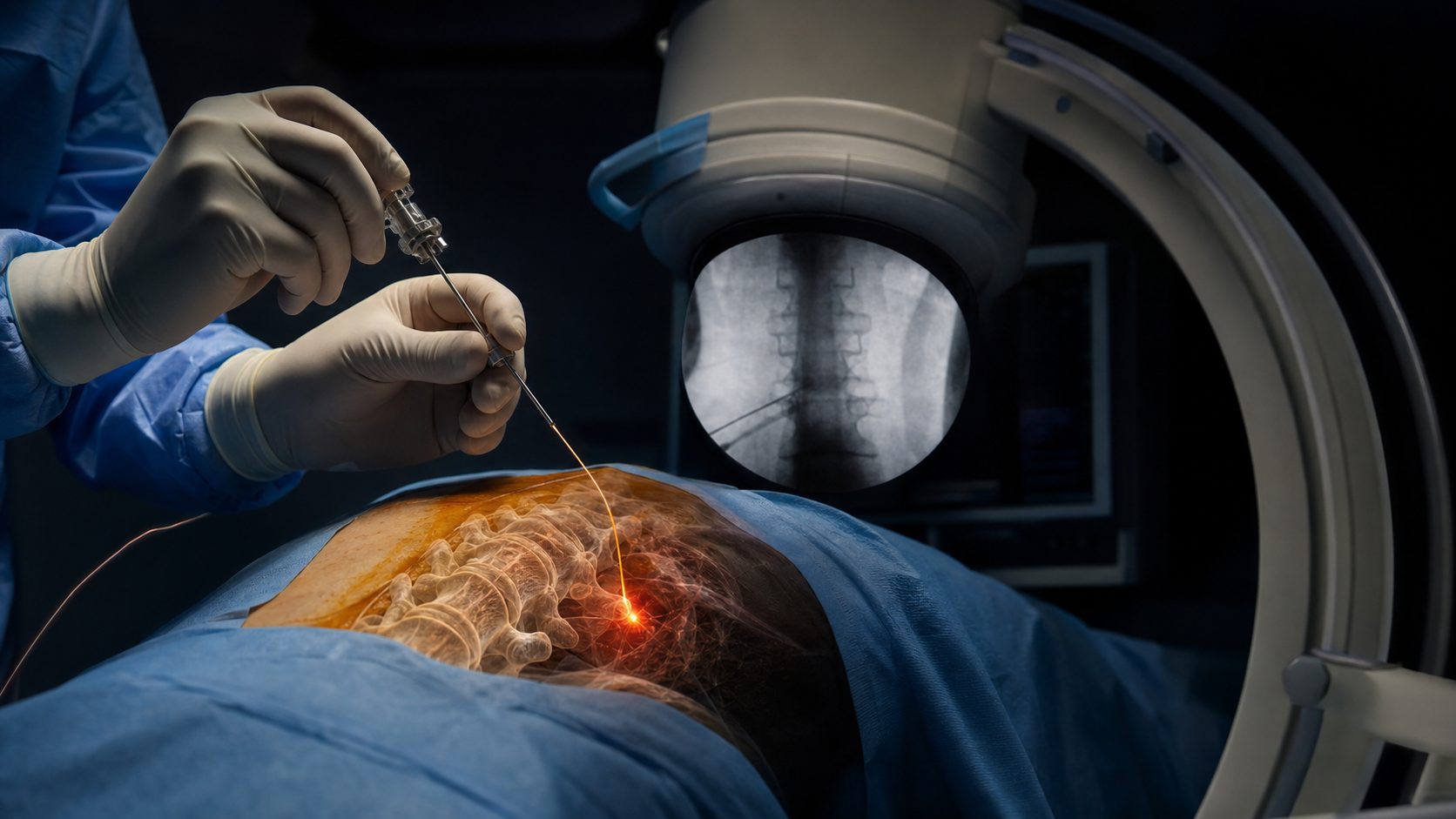
What Intradiscal Fractional Laser Actually Is
The Regenelase system is a 1550 nm erbium fiber laser that delivers sub-ablative (non-ablative) thermal energy through a 400 μm fiber-optic cable threaded through an 18-gauge spinal needle into the nucleus pulposus and inner annulus fibrosus under fluoroscopic guidance. This is not the cosmetic fractional laser used in dermatology — same wavelength family, entirely different clinical application and tissue target.
The critical regulatory fact: the Regenelase 1550 nm laser holds FDA 510(k) clearance K233344, and the cleared indications specifically include Spinal Surgery alongside General Surgery, Orthopedics, Podiatry, and Arthroscopy. The cleared indication covers surgical incision, excision, vaporization, ablation, and coagulation of soft tissue, including in conjunction with imaging. Device-level clearance for a surgical indication is a factual regulatory status — it is not a claim that any specific intradiscal reconstruction protocol is proven standard of care.
The procedural concept: the fiber delivers controlled photothermal energy at sub-ablative intensity (~60–70°C at the target zone), targeting the nucleus pulposus and inner annular fibers without removing tissue or altering gross disc architecture. The proposed mechanism involves three steps: thermo-mechanical micropore formation in the dense extracellular matrix, mechanotransduction signaling activating disc fibroblasts and chondrocyte-like nucleus pulposus cells, and neo-matrix formation over 6–12 weeks. The combination of multi-target PRP — delivered epidurally, intrafacetally, and into ligamentous structures in the same anesthetic encounter — is Pravida’s current combination protocol for complex discogenic presentations.
The Evidence: Baskov’s Series and What It Does and Doesn’t Tell Us
The Baskov 2015 J Spine Paper
The primary published citation for intradiscal fractional laser reconstruction is: Baskov AV, Borshchenko IA, Shekhter AB, et al. “Long Term Clinical Results in Laser Reconstruction of Spine Discs.” J Spine 4:210 (2015). Full text: OMICS Group direct URL; DOI: 10.4172/2165-7939.1000210.
The paper reports 97 patients with chronic discogenic back or neck pain treated with intradiscal non-ablative laser irradiation using a 1.56 μm Er:glass fiber laser, with five-year follow-up. The accessible published data describe: approximately 77% of patients showing MRI positive dynamics; approximately 95.9% VAS improvement; approximately 92% SF-36 improvement; no reported complications. Histologic findings in biopsied tissue described hyaline-like cartilage in the laser-treated zone of the nucleus pulposus.
What this is: an observational case series with encouraging signals and unusually long follow-up for this category of intervention. What this is not: a randomized sham-controlled trial. The absence of a control arm means we cannot attribute the improvement definitively to the laser procedure versus natural history, concurrent rehabilitation, regression to the mean, or patient selection. The fair evidence grade is preliminary signal-generating observational series — not proof of efficacy. A 2024 systematic review of image-guided minimally invasive options for degenerative lumbar spine disease (Ruffilli et al., Diagnostics 2024;14(11):1147) places percutaneous laser procedures in the broader minimally invasive treatment landscape without elevating them to standard-of-care status, which accurately reflects where the field currently sits.
The Longer Cohort: Sponsor-Reported Context
The Regenelase device sponsor (AcCELLerated Biologics) and CartiNova clinical education materials describe an ongoing 475-patient long-term experience under Baskov as principal investigator, dating from 2006 to the present, described as the longest-running fractional laser cartilage regeneration study in the world. We want to be transparent: this 475-patient framing comes from sponsor and clinical-deck materials — it is not a separately published peer-reviewed study we can independently verify. The distinction between a manufacturer-reported patient experience and a published peer-reviewed trial is clinically meaningful. We cite it here as context for the scope of institutional experience with this approach, not as equivalent evidence to the published 97-patient paper.
The Mechanism: Sobol et al. and the Biology of Sub-Ablative Laser in Avascular Tissue
The mechanistic science behind intradiscal laser reconstruction is supported by a body of work from Sobol and colleagues studying laser interactions with cartilage and disc tissue. The key publications: Sobol et al. J Biomed Opt 22(9):091515 (2017) and Sobol et al. J Biomed Opt 16(8):080902 (2011) describe laser-induced micropore formation and modification of cartilage extracellular matrix, along with mechano-transduction signaling cascades. A related paper — Alexandrovskaya et al. Laser Phys Lett 15:085601 (2018) — examines mechanisms of laser activation of chondrocytes in osteoarthritis healing. The proposed three-fold mechanism is: (1) thermo-mechanical micropore formation creates transient channels in the dense matrix, improving nutrient and metabolite exchange; (2) mechanotransduction signaling activates heat-shock proteins, growth factors, and chondrocyte clusters; (3) neo-matrix formation in the nucleus pulposus over subsequent weeks, including potential upregulation of aggrecan and type II collagen synthesis.
Biologic plausibility does not equal proven clinical benefit — but it does explain why this approach is worth investigating rigorously rather than dismissing outright. The mechanism is coherent with what we know about avascular tissue biology.
Why Discs Are Uniquely Hard to Regenerate
The intervertebral disc is the largest avascular tissue in the human body. The nucleus pulposus has a proteoglycan-to-collagen ratio of 27:1 — the highest of any tissue — with water content reaching 80–90% in youth, creating the hydrostatic pressure that absorbs spinal loading. Nutrition depends entirely on diffusion through the cartilaginous endplates; there is no blood supply to carry circulating repair cells into the disc interior. When nucleus pulposus cells degenerate, no cell type from outside the disc can replace the unique notochordal-derived cells that constitute the NP in early life.
This is why systemic biologics alone struggle to regenerate a disc: even if a biologic is delivered epidurally or intrathecally, penetrating the dense avascular matrix and reaching the NP is a fundamentally different challenge than treating a vascularized tissue. The hypothesis that controlled laser energy primes the disc for biological repair — by opening nano-pores in the matrix and triggering a local wound-healing response the tissue cannot otherwise mount — is grounded in this biology. Whether the clinical signal from Baskov’s series is the product of this mechanism or of other factors remains to be established in controlled trials.
Pravida Clinical Experience: Patient LB
At Pravida Health, we have begun offering intradiscal Regenelase for carefully selected patients. The following is a deidentified single-patient physician experience — one case, not a trial, and not generalizable.
Patient LB: 29-year-old male consultant with a four-year history of low back pain beginning with a tennis injury in December 2022. Severe flare three weeks prior to consultation. Imaging: L5–S1 degenerative disc disease with disc herniation into the endplate, S1 Modic edema, grade 1 anterolisthesis, left L5 nerve root closely approximating the disc. No leg numbness or tingling. Exam: pain on extension; flexion with hip-hinge guarding; left hip flexor TTP; negative straight-leg raise; lower extremity strength intact. No prior injections, surgery, or steroids; medication-averse.
Procedure performed 4/30/2026: intradiscal Regenelase 1550 nm at three sites × 50 seconds each via fiber-optic cable through spinal needle (collagenization of nucleus pulposus); intradiscal LR-PRP 2 cc; bilateral L5–S1 transforaminal LP-PRP 1.5 cc each (epidurography confirmed); intra-articular L5–S1 facet LR-PRP 1 cc each; fluoroscopy-guided platelet-poor plasma into L5 supraspinous, interspinous, and iliolumbar ligaments and L5 multifidi. Antibiotic prophylaxis was administered for intradiscal access (standard practice given the approximately 1% risk of discitis without prophylaxis in intradiscal procedures). Total platelet delivery across all sites: approximately 7.9 billion platelets. Expected improvement window: 6–12 weeks post-procedure. Activity reintroduction at 6 weeks; MRI re-evaluation if symptoms unresolved at 12 weeks. This was a single-anesthetic encounter addressing multiple pain generators simultaneously.
“A controlled photothermal injury inside an avascular disc is an entirely different proposition from injecting a biologic on top of one. Whether that matters clinically — durably, across patients — is still being characterized.”
How We Use This at Pravida Health
Pravida Health is located at 1801 Peachtree St NE, Ste 150, Atlanta, GA 30309. Intradiscal Regenelase is offered as part of our CartiNova regenerative spine service line — a broader framework that includes the CartiNova BioProfile™ whole genome sequencing clinical decision framework, orthobiologic combinations (PRP, BMAC), and phenotype-guided rehabilitation. The laser is not a standalone solution; it is a matrix primer in a multi-component strategy.
Who Is a Candidate
Patient selection for intradiscal laser requires all of the following: a confirmed discogenic pain phenotype (not facet-mediated, not SI-joint, not primarily radicular with structural compression); demonstrated failure of conservative care including targeted physical therapy, graded activity, sleep and load optimization, and anti-inflammatory strategy; absence of progressive neurologic deficit; absence of major structural instability requiring surgical stabilization; and realistic expectations about uncertain benefit. This is not appropriate for patients with significant nerve compression who need surgery, and it is not appropriate as a first-line treatment before conservative care has been genuinely optimized.
The Combination Protocol
Our current protocol is: intradiscal laser first — to prime the matrix and create nano-pores that improve biologic penetration — followed immediately by intradiscal PRP or BMAC delivery, with concurrent treatment of adjacent pain generators (facet joints, epidural space, paraspinal ligaments) in the same anesthetic encounter where indicated. Antibiotic prophylaxis is standard for intradiscal access given the approximately 1% discitis risk documented in standard intradiscal procedure literature. Contact us if you would like to discuss whether this protocol is appropriate for your specific diagnosis.
Risks, Limitations & What the Evidence Doesn’t Show
We do not want the clinical excitement around a novel device to obscure what the evidence actually supports. The following limitations are important:
- Discitis risk: Any intradiscal procedure carries a risk of disc space infection (discitis) of approximately 1% without antibiotic prophylaxis. We administer prophylactic antibiotics for every intradiscal access. Patients should understand this risk and discuss it fully before consenting.
- Bleeding and nerve irritation: As with any image-guided spinal procedure, there is a risk of bleeding, nerve root irritation, and transient worsening of symptoms in the post-procedure period.
- MRI changes ≠ symptom improvement: Baskov’s series reported MRI positive dynamics in 77% of patients. Imaging change and meaningful clinical improvement are not the same outcome. A disc that appears different on MRI may still generate pain.
- Observational series ≠ RCT: The published Baskov paper is an observational case series, not a randomized sham-controlled trial. We cannot confidently attribute benefit to the laser procedure itself.
- No sham-controlled blinded trial yet: As of this writing, there is no published randomized, blinded, sham-controlled trial of intradiscal fractional laser reconstruction. This is the most important evidence gap.
- Not a substitute for surgery: Progressive neurologic deficit, significant structural nerve compression, or spinal instability requiring stabilization are surgical indications. Delaying definitive surgery to pursue investigational laser treatment in these patients can result in irreversible neurologic injury. This point is non-negotiable.
- Sponsor-reported vs. peer-reviewed data: The 475-patient cohort framing comes from sponsor materials. Manufacturer-reported clinical experience is not equivalent to independent peer-reviewed evidence, and we flag this distinction explicitly.
What You Can Do Today
- Confirm the pain generator. Disc degeneration on MRI does not automatically mean the disc is causing your pain. A structured evaluation distinguishing discogenic pain from facet-mediated pain, SI-joint pain, radicular pain, and myofascial deconditioning is the essential first step. Procedures only work when applied to the correct diagnosis.
- Optimize conservative care genuinely before pursuing procedures. Targeted physical therapy, graded loading, sleep quality, anti-inflammatory nutrition, and weight management have far deeper evidence bases than any spine injection or laser procedure. These are not consolation prizes — they are prerequisites. Contact Pravida Health to speak with a physician about what a complete conservative care program should include for your diagnosis.
- Get a structured imaging review. MRI severity and symptom severity often diverge substantially. A board-certified spine physician reading the images in context of your clinical presentation can tell you whether the imaging finding is likely the pain generator — or whether the disc is an incidental finding.
- Ask about evidence grade directly. If a clinic proposes intradiscal laser, ask: Is there a randomized sham-controlled trial? What is the evidence grade for this specific protocol? What are the realistic risks? A physician who cannot answer those questions clearly is a concerning sign.
Frequently Asked Questions
Is the Regenelase device FDA-cleared for spinal use?
Yes. The Regenelase 1550 nm Er fiber laser holds FDA 510(k) clearance K233344, which specifically lists Spinal Surgery among its cleared indications alongside General Surgery, Orthopedics, Podiatry, and Arthroscopy. Device-level clearance for a surgical use category is a regulatory fact. It does not constitute FDA approval of a specific clinical reconstruction protocol, nor does it mean intradiscal fractional laser is standard of care for degenerative disc disease. Cleared device and proven clinical efficacy for a specific indication are two separate questions, and we distinguish between them carefully.
What did Baskov’s study actually show?
Baskov et al. J Spine 2015 (DOI: 10.4172/2165-7939.1000210) reported a 97-patient observational series of intradiscal non-ablative laser irradiation with 1.56 μm Er:glass fiber laser and 5-year follow-up. Published data describe approximately 77% MRI positive dynamics, 95.9% VAS improvement, and 92% SF-36 improvement, with no reported complications. This is an encouraging clinical signal from an unusually long follow-up series. It is not a randomized sham-controlled trial, so findings cannot be definitively attributed to the laser procedure itself versus natural history or other concurrent treatments. Read it as a well-documented observational series generating a hypothesis worth testing in controlled trials.
How is this different from disc surgery or microdiscectomy?
Conventional microdiscectomy removes herniated disc material to decompress a nerve root — it is a decompressive procedure for structural nerve compression. Intradiscal fractional laser targets the nucleus pulposus and inner annulus with sub-ablative thermal energy to stimulate matrix remodeling, not to remove tissue. The two procedures address different clinical problems: surgery is appropriate for structural nerve compression with neurologic deficit; intradiscal laser is being investigated for discogenic pain in patients without surgical indications. They are not interchangeable, and laser is not a substitute when surgery is clearly indicated.
Can intradiscal laser regenerate a disc?
The biologic hypothesis is that controlled photothermal injury inside the nucleus pulposus can trigger mechanotransduction signaling, nano-pore formation in the dense avascular matrix, and neo-matrix synthesis — a repair response the disc cannot mount on its own due to its complete avascularity. Baskov’s histologic data described hyaline-like tissue in the treated zone. Whether this translates to durable disc regeneration that outperforms sham or natural history in a randomized trial remains unproven. The disc can change on MRI without the patient meaningfully improving — and vice versa. The honest answer: possibly, under carefully selected conditions, but this has not been established in a controlled trial.
Is this offered at Pravida Health?
Yes. Pravida Health at 1801 Peachtree St NE, Ste 150, Atlanta, GA 30309 offers intradiscal Regenelase as part of the CartiNova regenerative spine service line for carefully selected patients. Selection requires a confirmed discogenic phenotype, failure of conservative care, absence of progressive neurologic deficit or major structural instability, and a frank discussion about the current evidence grade. We do not frame this as a proven treatment — we frame it as an evidence-informed option with a biologically sound mechanism and an encouraging but not definitive clinical signal. Contact us to schedule a consultation and discuss whether this is appropriate for your specific situation.
Want a physician-level evidence review of your spine options?
At Pravida Health in Atlanta, we start with your diagnosis, your imaging, and what the evidence actually supports for your specific pain generator — not a preferred device or procedure. If intradiscal laser is appropriate for your case, we will tell you. If surgery, rehabilitation, or a different regenerative approach is the better fit, we will tell you that too.
Schedule a Consultation