Knee osteoarthritis robs patients of walking tolerance, exercise capacity, and the ability to live around their goals rather than around their pain. When patients hear the word “laser” applied to the knee, two responses are equally predictable: uncritical enthusiasm or reflexive skepticism. Neither serves them well. The honest answer sits in between — and it requires distinguishing what is a regulatory fact, what is preclinical biology, what is case-level clinical evidence, and what remains unknown.
The Regenalase system is a real device with FDA 510(k) clearance. The preclinical histology showing hyaline cartilage regeneration in mini-pig models is published peer-reviewed science. The Scarpone case series is a set of physician case reports — not a randomized trial — that produced signals worth taking seriously. And our own deidentified experience at Pravida Health includes an outcome that was striking enough to present at a clinical forum. This article lays all of that out, evidence-graded, with appropriate caveats at each level. The fundamentals — physical therapy, weight optimization, load management, anti-inflammatory care — still come first. The laser is a next-step option for carefully selected patients, not a first-line treatment and not a substitute for joint replacement when that is truly what is needed.
What Fractional Laser Treatment for Cartilage Actually Is
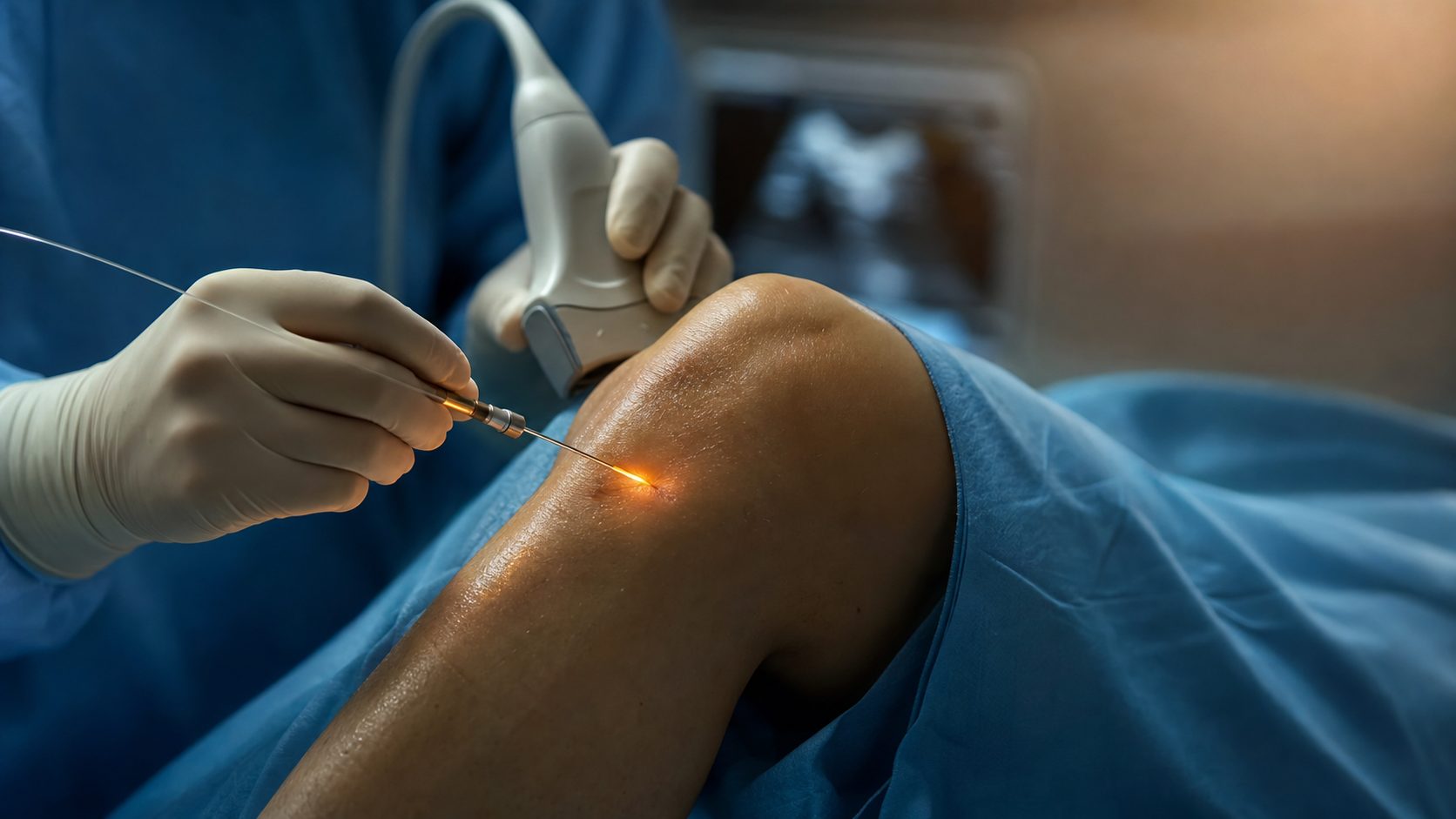
The Regenalase system is a dual-laser platform. The primary component is a 1550 nm erbium fiber laser delivered through a 400 µm optical fiber threaded through an 18-gauge cannula. This enables minimally invasive, percutaneous access to cartilage and other soft-tissue joint structures under ultrasound or nanoscope guidance — no open incision, no general anesthesia. The secondary component is a 980 nm diode laser providing non-invasive photobiomodulation (PBM) at the surface, activating cytochrome c oxidase to increase ATP production, reduce inflammation, and augment the effect of the interstitial laser work.
The device holds FDA 510(k) clearance K233344, which covers vaporization, incision, excision, ablation, cutting, hemostasis, and coagulation of soft tissue including meniscus cartilage, in conjunction with imaging, across the specialties of General Surgery, Orthopedics, Podiatry, Arthroscopy, and Spinal Surgery. The 980 nm component is separately cleared for temporary pain relief, muscle spasm reduction, and increased local circulation. Clearance can be verified through the FDA 510(k) database. That clearance speaks to device safety and engineering. It does not, by itself, establish efficacy for cartilage regeneration or osteoarthritis disease modification — that is what the clinical and preclinical evidence addresses.
The procedure is typically office-based under local lidocaine, lasting approximately 30 minutes. The 18-gauge needle approach means a similar access risk profile to a diagnostic joint aspiration. Patients generally return to normal activity within 24–48 hours of the procedure.
The Evidence: What This Laser Can and Cannot Do Today
FDA Clearance: What It Covers and What It Does Not
FDA 510(k) clearance is a device-level determination that a new device is substantially equivalent in safety and effectiveness to a predicate device already on the market. It is not an efficacy determination for a specific clinical indication — it is not the same as FDA approval of a drug therapy for knee osteoarthritis. The clearance for K233344 permits soft-tissue surgical use including cartilage procedures in conjunction with imaging. Physicians may use cleared devices in their clinical judgment within and around cleared indications. Any claim that the laser “treats osteoarthritis” or “regenerates cartilage” as a defined therapeutic indication goes beyond what the clearance establishes — that is the clinical evidence question, addressed below.
Mechanism: The Three-Step Model
The biological rationale for fractional laser treatment in avascular tissue like articular cartilage follows a coherent three-step model. First, heating response induction: sub-ablative thermal stress at approximately 60–70°C creates microscopic zones of coagulation inside cartilage without surface incision, triggering a localized wound-healing cascade in tissue that otherwise has no intrinsic repair capacity. Second, nano-pore formation in the ECM: laser-induced gas vacuoles open transient channels in the dense extracellular matrix, enabling nutrient and metabolite exchange that the avascular tissue cannot achieve on its own — and enabling penetration of subsequently delivered biologics. Third, mechano-transduction signaling: the thermomechanical stresses activate chondrocyte mechanoreceptors, upregulating heat-shock proteins, growth factors, and collagen II and proteoglycan synthesis. The net effect, if the preclinical and case-level evidence is directionally correct, is a wound-healing microenvironment that avascular cartilage cannot generate without external stimulus.
Preclinical Evidence: Mini-Pig Hyaline Cartilage Regeneration
The most compelling basic-science evidence comes from animal studies published in peer-reviewed optics and biomedical journals. In a dual-defect mini-pig model, cartilage defects were created on Day 1; one defect was treated with the fractional laser, the other served as an untreated control. By Day 60, the treated side showed measurable repair activity. At Day 120 histology, the treated defects showed hyaline cartilage coverage — not fibrocartilage scar — while untreated controls did not. This is the distinction that matters clinically: hyaline cartilage (type II collagen, proper biomechanical properties) versus fibrocartilage (inferior repair tissue from microfracture approaches). These findings are reported in Sobol et al., Journal of Biomedical Optics 22(9) 091515 (2017) and corroborated by chondrocyte activation mechanism data in Alexandrovskaya et al., Laser Physics Letters 15(8) 085601 (2018). Earlier foundational work on laser-induced cartilage regeneration was described in Sobol et al., Journal of Biomedical Optics 16(8) 080902 (2011). These are preclinical findings — they establish biological plausibility and directional evidence, not clinical proof.
Scarpone Case Series: Patients EL, MB, and BM
Dr. Michael Scarpone, a sports medicine physician, reported three cases of Stage 3–4 knee osteoarthritis treated with the Regenalase fractional laser combined with intra-articular and intraosseous BMAC (bone marrow aspirate concentrate). These are physician case reports from outpatient practice — not a blinded, controlled trial — and should be interpreted accordingly.
Patient EL was a 62-year-old with BMI 34, Stage 3–4 OA, and an unrepaired ACL. Imaging showed bone marrow edema and cartilage loss at the medial femoral condyle. She had been using daily NSAIDs, had received cortisone and hyaluronic acid injections, and had been recommended a total knee replacement by Hospital for Special Surgery. Following treatment with BMAC (intra-articular plus intraosseous) combined with the fractional laser, she achieved resolution of marrow edema, improved cartilage thickness in the medial compartment on 6-month MRI, joint space increase, and complete discontinuation of all pain medications. She reported daily activity and sleep without pain. A confirmatory finding: the untreated compartment (Stage 1 OA) showed no change — consistent with treatment specificity rather than a systemic or placebo effect.
Patient MB was a 52-year-old with BMI 24, Stage 3–4 OA, and prior right total knee replacement. The treated left knee had undergone prior ACL reconstruction and meniscectomy, with subchondral bone swelling and cartilage loss on imaging. She had been using nearly daily NSAIDs and received a knee replacement recommendation from Lahey Clinic. Post-treatment outcomes included resolution of marrow edema, improved cartilage thickness at the tibial plateau, elimination of pain medications and knee brace, and full return to daily activities and pain-free sleep. The significance here is that a meaningful response occurred in a post-surgical knee with prior tissue excision — a harder environment than native tissue.
Patient BM was a 57-year-old with BMI 29, Stage 3 OA, and femoral condyle involvement. She had tried prior PRP, bone marrow injections, and cortisone without durable relief and was experiencing increasing pain with daily activity. Post-treatment outcomes included resolution of marrow edema, improved cartilage thickness, reduced pain medication use, and improved daily activity and sleep quality. This case is notable because the patient had failed prior biologic interventions — consistent with the hypothesis that laser tissue preparation is the differentiating variable.
These are case reports, not a randomized controlled trial. They do not establish causality. Placebo effects, natural history, and regression to the mean cannot be fully excluded. But three cases across independent physicians, all with objectively measured MRI changes, all with Stage 3–4 OA, and one with a within-patient control (treated vs. untreated compartment), represent a meaningful clinical signal that justifies ongoing prospective study.
Pravida Clinical Experience: Patient JA
Our own deidentified case was presented at the TOBI Networks Clinical Forum in May 2026. Patient JA is a 58-year-old female endurance runner with bilateral knee OA diagnosed more than 10 years prior — she had stopped playing tennis because of her knees but continued competing in distance running with 17+ marathons and 129 half-marathons completed. She presented in December 2025 with right knee pain that had been refractory to coaching modifications, physical therapy, and brace intervention. X-ray showed mild medial joint line narrowing and small bilateral femoral condyle osteophytes.
She underwent a single 30-minute office procedure under local lidocaine: Regenalase 1550 nm interstitial laser monotherapy (no PRP, no BMAC) under ultrasound guidance, treating approximately nine cartilage sites along the medial femoral cartilage interface. Within the first 24 hours, she reported minimal pain — well below her expected baseline. At three weeks, she completed her first 10-mile endurance run pain-free. At four weeks, she completed a half-marathon with zero pain, slightly off race pace, and eliminated her running brace entirely.
This is a single deidentified case. It is not a controlled trial and results cannot be generalized. The outcome is striking enough to report — high-mileage athlete, refractory to prior conservative care, single-modality laser treatment, functional restoration within one month — but it requires prospective study to determine how reproducible it is, in which patient profiles, and with what durability. We report it because transparency about real-world clinical experience is part of how physicians honestly counsel patients.
“FDA clearance speaks to safety and device-level engineering. Clinical efficacy in cartilage regeneration is still being characterized — and that distinction has to drive how we counsel patients.”
How We Use This at Pravida Health
The Regenalase fractional laser is offered within our CartiNova regenerative orthopedics service line at Pravida Health, located at 1801 Peachtree St NE, Ste 150, Atlanta, GA 30309. CartiNova integrates the laser with a genomic decision framework (the CartiNova BioProfile™, based on whole-genome sequencing) to individualize biologic selection, prehabilitation, and rehabilitation protocols. The laser is a tissue primer; the biologic (BMAC, PRP, or MFAT selected by genomic phenotype) is the regenerative payload delivered into laser-prepared channels.
Patient Selection
We are deliberate about who is and is not an appropriate candidate. The patients we evaluate seriously are those with Stage 3–4 knee osteoarthritis that is refractory to a structured conservative care program — meaning they have genuinely attempted physical therapy, weight optimization where relevant, appropriate load management, and anti-inflammatory strategies. We exclude patients with severe mechanical deformity, significant varus or valgus alignment requiring corrective osteotomy, or truly end-stage bone-on-bone disease where joint replacement is the medically appropriate next step. Laser-based regenerative care is not a substitute for total knee replacement when that is what a patient actually needs — using it in that context delays appropriate care.
Combination with BMAC
For most candidates, we combine the laser with intra-articular and intraosseous BMAC. The rationale is that laser nano-pore formation opens the ECM so that BMAC-delivered mesenchymal stem cells can penetrate the avascular tissue and access the chondrocyte signaling environment created by the laser. The ongoing US clinical trial NCT04001361 is investigating this combination (laser plus bone marrow grafting) in knee cartilage defects with n=52 enrolled patients. Early data from that trial, as well as from the completed Baskov knee study (n=12, statistically significant VAS and SF-36 improvement at 12 months), support the biological rationale. We use the CartiNova BioProfile™ genomic phenotype to determine whether BMAC, PRP, or MFAT is the appropriate biologic complement for each patient.
What We Measure
We do not treat without an outcomes framework. Before and after the procedure, we track: KOOS (Knee injury and Osteoarthritis Outcome Score), VAS pain scores, walking tolerance, stair tolerance, sleep disruption, and medication use. MRI is obtained at baseline and at 6 months when clinically indicated to evaluate marrow edema resolution and any cartilage changes. If a treatment cannot move those measures in a meaningful and durable direction for a given patient, we need to know that — and the patient deserves to know it too.
Risks, Limitations & What the Evidence Doesn’t Show
Honest patient counseling requires being as specific about what we do not know as about what we do.
- Thermal effects and tissue injury: Sub-ablative laser energy is controlled by design, but imprecise targeting or excessive energy delivery could cause unintended thermal injury to surrounding structures. Experienced proceduralists with imaging guidance reduce but do not eliminate this risk.
- Infection: Any percutaneous joint access carries infection risk. For Regenalase procedures, this is a low but real risk, mitigated by sterile technique and appropriate patient selection. Intradiscal procedures require antibiotic prophylaxis; knee procedures carry a lower baseline infection risk than disc access.
- No high-quality multi-center RCT yet: The absence of a published randomized controlled trial is the most important limitation. The Scarpone cases and our own clinical experience are signal, not proof. NCT04001361 is ongoing; its results will be important. Until controlled data exist, the evidence grade is preclinical plus case-level.
- Scarpone reports are case-level, not blinded: Physician case reports cannot exclude placebo, natural history, concurrent lifestyle changes, or regression to the mean. The within-patient control in Patient EL’s case (untreated compartment unchanged) is the most rigorous internal check, but it is not a substitute for a controlled trial design.
- MRI changes do not always equal durable pain relief: Marrow edema resolution and cartilage thickening on MRI are objective findings. But structural MRI changes and patient-reported pain outcomes do not always track together, and the durability of MRI improvements at longer follow-up (beyond 6–12 months) is not yet established for this approach.
- Not a substitute for joint replacement when truly indicated: When a patient has end-stage OA with major bone-on-bone deformity, mechanical instability, or failed multiple prior interventions, joint replacement is the appropriate recommendation. Using laser-based regenerative care to delay surgery in those patients does them harm, not good.
- Individual results vary: Outcomes in the Scarpone series and at Pravida Health reflect individual cases. Patient selection, disease severity, prior treatment history, body weight, alignment, and biological response all influence outcomes. No outcome can be guaranteed.
What You Can Do Today
- Confirm the diagnosis before pursuing any treatment. Knee pain is not always osteoarthritis. Meniscal pathology, inflammatory arthritis, referred pain from the hip or spine, and tendinopathy can all mimic OA. Imaging and clinical examination by a physician with specific musculoskeletal expertise are the starting point.
- Exhaust the proven conservative care program first. A structured quadriceps and hip-strengthening program, gait optimization, appropriate footwear, weight management when relevant, and selective use of topical or oral NSAIDs constitute the evidence base that any regenerative option must be compared against. These are not consolation prizes — they are required prerequisites.
- Get a proper imaging review. Weight-bearing X-rays and, where indicated, MRI with attention to marrow edema patterns help define disease severity, alignment, and whether the structural picture is compatible with a regenerative approach or is more appropriately addressed by replacement surgery.
- Ask your physician to grade the evidence for any proposed intervention. FDA clearance, preclinical animal data, physician case series, and randomized controlled trials represent different evidence levels and should be described to you as such. If a clinician presents case reports as if they were RCT-level evidence, that is a red flag.
- Demand specific outcomes data. Ask what measures will be tracked, how long follow-up extends, and what the threshold for success is before the procedure. Contact us at Pravida Health if you would like a structured evaluation of whether this approach fits your specific situation.
Frequently Asked Questions
Is Regenalase FDA-approved for treating knee osteoarthritis?
The Regenalase system holds FDA 510(k) clearance K233344 for vaporization, incision, excision, ablation, and coagulation of soft tissue including meniscus cartilage, in conjunction with imaging. That is a device-level clearance for soft-tissue surgical procedures — it does not constitute an FDA-approved indication specifically for osteoarthritis treatment or cartilage regeneration as a defined clinical claim. Clinical use within cleared soft-tissue indications is permitted; cartilage regeneration or OA disease modification remains investigational and is the subject of ongoing prospective study.
What did Dr. Scarpone’s cases actually show?
Dr. Michael Scarpone reported three cases — Patients EL, MB, and BM — all with Stage 3–4 knee osteoarthritis treated with Regenalase laser combined with intra-articular and intraosseous BMAC. Each case showed resolution of marrow edema, improved cartilage thickness on MRI follow-up, and meaningful reductions in pain medication use. One case (Patient EL) had a within-patient control showing no change in the untreated compartment. These are physician case reports from outpatient practice, not a controlled trial. They are meaningful clinical signals, but they do not establish causality and results cannot be generalized to other patients without prospective controlled data.
How is this different from PRP or BMAC alone?
Articular cartilage is avascular — its dense extracellular matrix limits diffusion of growth factors and there is no blood supply to recruit circulating repair cells. PRP and BMAC deliver bioactive signals, but those signals have limited penetration into intact, degenerated cartilage without tissue preparation. The Regenalase laser creates microscopic pores in the ECM, induces a localized wound-healing response, and activates chondrocyte mechanoreceptors — creating the microenvironment that allows BMAC-delivered mesenchymal stem cells to differentiate toward a chondrocytic rather than fibroblastic phenotype. Patient BM in the Scarpone series, who had already failed prior PRP and BMAC injections, responded after the addition of laser tissue preparation — the most direct clinical illustration of this hypothesis.
Can the laser regenerate cartilage?
Mini-pig preclinical studies by Sobol et al. (J Biomed Opt, 2017) demonstrated histologic evidence of hyaline cartilage coverage in laser-treated defects at 120-day follow-up, compared with untreated controls. In the Scarpone case series, treated patients showed MRI cartilage thickening and marrow edema resolution. MRI changes, however, do not always translate to durable long-term pain relief, and no multi-center randomized controlled trial has yet confirmed cartilage regeneration as a reproducible clinical outcome. The evidence grade is preclinical plus case-level — biologically compelling, clinically promising, not yet proven by RCT standards.
Is this offered at Pravida Health?
Yes. The Regenalase 1550 nm fractional laser is offered within the CartiNova regenerative orthopedics service line at Pravida Health, 1801 Peachtree St NE, Ste 150, Atlanta, GA 30309. Patient selection is careful: we focus on Stage 3–4 knee OA refractory to conservative care, without severe deformity or mechanical instability requiring joint replacement. We track outcomes with KOOS, VAS pain scores, walking tolerance, and MRI at follow-up. Contact us to determine whether you are an appropriate candidate.
Discuss whether you’re a candidate
At Pravida Health, we start with your diagnosis, your imaging, and your treatment history — not a preferred procedure. If the Regenalase laser and CartiNova protocol make sense for your situation, we will tell you. If the fundamentals should come first, or if joint replacement is the right answer, we will tell you that too.
Request an Evaluation