A patient came to Pravida Health after spending three weeks calling Atlanta imaging centers trying to find a place that offered DEXA with Trabecular Bone Score analysis. She had been diagnosed with osteopenia two years earlier and felt increasingly uneasy about a treatment decision her referring physician described as a “close call.” Her T-score sat at −2.1 — firmly in the osteopenic range, near the boundary where the calculus around pharmacologic treatment starts to matter. She was not sure whether to start bisphosphonate therapy. Neither, honestly, was her doctor.
What she did not know — what her imaging center had not offered — was that her standard DEXA result was only telling half the story. The T-score measures bone density: how much mineral is packed into a cubic centimeter of bone. What it does not measure is bone architecture: how that mineral is organized inside the trabecular network. Two patients with identical T-scores can have dramatically different fracture risk because the microarchitecture of their bone is different. Trabecular Bone Score (TBS) is the tool that closes this gap — and it adds zero additional radiation, zero additional scan time, and requires no separate office visit. It is a software re-analysis of the lumbar spine DEXA image you already have.
What a Standard DEXA Tells You — and What It Misses
Think of bone as a structural material rather than a uniform solid. Imagine two wooden beams of identical density — same weight per cubic foot. One is old-growth oak with tight, regular grain. The other is pine with wide, irregular grain full of micro-gaps. Both weigh the same. The oak holds the load; the pine fails. The DEXA T-score is the weight measurement. It tells you density. It cannot tell you about the grain.
Trabecular bone — the spongy, lattice-like interior of vertebral bodies and femoral necks — is the structural material most relevant to osteoporotic fracture. Vertebral compression fractures and hip fractures happen when this lattice fails under load. The quality of that lattice — the connectivity, spacing, and regularity of the trabecular rods and plates — is what TBS measures. A bone with excellent density but degraded microarchitecture is exactly the bone that surprises a patient with a fracture after a minor fall. It is also the bone that a standard DEXA report describes as “low-normal” while missing the real structural problem.
“Density isn’t strength. Two patients with identical T-scores can have dramatically different fracture risk — because the microarchitecture of the bone is different.”
This gap is not hypothetical. Silva et al. (Bone, 2015) established that TBS independently predicts fracture risk beyond what T-score and FRAX alone capture. The association with vertebral and hip fracture held across major population cohorts even after adjusting for BMD and clinical risk factors. TBS adds signal that BMD simply does not contain.
What TBS Actually Measures
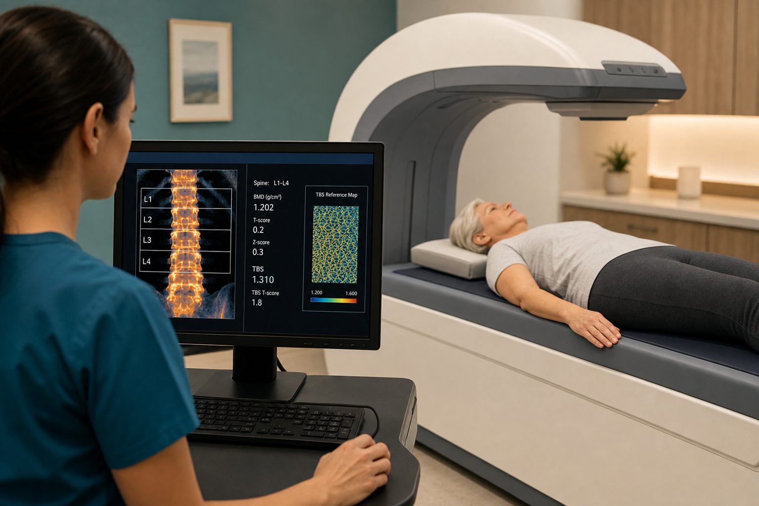
TBS is calculated by the Medimaps TBS iNsight platform, applied as a software layer on top of the DXA image acquired during standard lumbar spine scanning. The algorithm analyzes the gray-level texture of the L1–L4 region of interest — the same vertebral bodies captured in a standard DEXA — and extracts a quantitative index of trabecular microarchitecture. Higher values indicate a better-connected, more fracture-resistant lattice. Lower values indicate degraded microarchitecture with increased fracture vulnerability.
The critical clinical implication is that TBS integrates into the FRAX algorithm as a multiplicative adjustment. A patient with a borderline FRAX score and a low TBS gets their 10-year fracture probability adjusted upward — which can push a “close call” patient clearly into treatment territory. The reverse is also true: a patient with a moderately low BMD but well-preserved microarchitecture may have a lower true fracture risk than their T-score alone suggests.
What the Evidence Says
Four high-quality publications anchor the clinical case for TBS:
- Silva et al., Bone 2015 (PMC4538791) — established TBS as an independent predictor of osteoporotic fracture across major population cohorts, holding after adjustment for BMD and standard FRAX inputs. This is the foundational validation study that put TBS on the clinical map.
- Florescu et al., J Med Life 2020 (PMC7803323) — confirmed TBS utility in secondary osteoporosis populations including patients with type 2 diabetes, glucocorticoid use, and hyperparathyroidism, where standard BMD is particularly likely to underestimate true fracture risk.
- ISCD 2023 Adult Positions PDF (iscd.org) — the International Society for Clinical Densitometry formally recommended TBS analysis in all adults aged 40 and older undergoing lumbar spine DXA, encoding TBS as standard-of-care rather than investigational.
- Shevroja et al., J Clin Densitom 2024 (ScienceDirect) — the 2023 ISCD Official Positions paper quantified the reclassification impact: TBS-adjusted FRAX changes clinical management in approximately 9–18% of patients who sit close to accepted intervention thresholds.
That last finding deserves its own emphasis:
“TBS-adjusted FRAX changes clinical management in approximately 9–18% of patients who sit close to accepted intervention thresholds — exactly the people where the right call matters most.”
Who Benefits Most From Adding TBS
TBS adds value across a wide range of patients, but its reclassification impact is highest in six populations:
- Patients sitting on the treatment fence — FRAX scores near the intervention threshold where TBS-adjusted FRAX changes the clinical decision in 9–18% of cases (Shevroja et al. 2024).
- Type 2 diabetes — patients with T2D often have normal or near-normal BMD despite significantly degraded trabecular microarchitecture. Glycation of collagen and other structural proteins impairs bone quality in ways that density measurement misses (Florescu et al. 2020).
- Long-term glucocorticoid use — glucocorticoids preferentially damage trabecular architecture, often before significant BMD change is apparent on DEXA. TBS detects this structural degradation early (Florescu et al. 2020).
- Hyperparathyroidism and other secondary causes — parathyroid hormone excess causes cortical bone loss but also trabecular disruption; TBS captures microarchitectural changes that precede or exceed BMD-measurable bone loss.
- Postmenopausal women with normal BMD but elevated clinical risk — a normal T-score in the setting of elevated fall risk, prior fragility fracture, or strong family history may be falsely reassuring if microarchitecture is not assessed (Silva et al. 2015).
- Men over 50 — osteoporosis in men is underdiagnosed and undertreated; TBS provides additional fracture risk stratification in a population where standard DEXA thresholds have less validation.
What a TBS-Enhanced Evaluation Looks Like at Pravida Health
When you come to Pravida Health in Atlanta for a DEXA + TBS evaluation, the visit produces a substantially more complete picture than a standard bone density scan. Our evaluation includes:
- Full BMD report — standard lumbar spine and femoral neck T-scores with standard interpretation.
- TBS analysis of L1–L4 using the Medimaps TBS iNsight platform — applied to the same image, no additional radiation or scan time.
- TBS-adjusted FRAX 10-year fracture risk — major osteoporotic fracture and hip fracture probabilities recalculated with TBS as a FRAX modifier.
- Written summary for patient and referring physician — a clear report explaining both the BMD and TBS findings in clinical context, including what the combined picture means for treatment decision-making.
- Optional regenerative medicine and bone-health consult — for patients who want to discuss non-pharmacologic and precision-medicine approaches to bone health alongside the standard treatment conversation. Book a consultation to discuss your options.
A Better Conversation, Not Just a Better Number
The patient who called around Atlanta imaging centers and came to us with her “close call” osteopenia diagnosis got her TBS result at the same appointment as her DEXA. Her TBS was 1.185 — below the 1.230 degraded-microarchitecture threshold. Her TBS-adjusted FRAX pushed her clearly across the intervention threshold. The ambiguity was resolved. The clinical decision was no longer a close call. That is what a complete evaluation looks like.
The hard truth is that most Atlanta imaging centers still offer standard DEXA without TBS analysis. The data is in the image; the software is what is missing. If you have been evaluated for osteoporosis, have a borderline T-score or FRAX score, or have a condition that makes standard BMD likely to underestimate your fracture risk — a TBS-enhanced evaluation is worth a conversation.
Ready for a complete bone health evaluation?
Pravida Health offers DEXA scanning with full Trabecular Bone Score analysis in Atlanta — same scan, richer picture, clearer decision. We serve patients at 1801 Peachtree St NE, Ste 150, Atlanta, GA 30309.
Book a DEXA + TBS EvaluationCall 404.900.7371 · info@pravida.com
Frequently Asked Questions
What is the Trabecular Bone Score (TBS)?
TBS is a software-based analysis of the lumbar spine DEXA image that quantifies bone microarchitecture — specifically the texture and connectivity of the trabecular network in L1–L4. Unlike BMD, which measures how much mineral is present, TBS reflects how well that mineral is organized. A high TBS indicates a well-connected, fracture-resistant microarchitecture; a low TBS (<1.230) indicates degraded, fracture-prone trabecular structure (Silva et al., Bone 2015). TBS is calculated by Medimaps TBS iNsight software applied to the existing DEXA image with no additional radiation or scan time.
Does TBS require a separate scan or additional radiation?
No. TBS is a software re-analysis of the lumbar spine DEXA image you already had. It adds zero additional radiation, zero additional scan time, and requires no separate office visit. The Medimaps TBS iNsight platform applies a texture analysis algorithm to the L1–L4 region of interest in the existing DXA image and generates a TBS score alongside the standard BMD and T-score report. This is confirmed in the 2023 ISCD Official Positions.
Who should get a TBS-enhanced DEXA?
TBS is most clinically useful for patients near the FRAX treatment threshold, patients with type 2 diabetes, those on long-term glucocorticoids, patients with hyperparathyroidism or other secondary causes of bone loss, postmenopausal women with normal or osteopenic BMD but elevated clinical risk, and men over 50 with risk factors. The ISCD formally recommends TBS in all adults aged 40+ undergoing lumbar spine DEXA (ISCD 2023 Official Positions). See also Florescu et al. 2020 for secondary osteoporosis populations.
How does TBS change my fracture risk score?
TBS is integrated into the FRAX algorithm as a multiplicative adjustment to the 10-year fracture probability. A low TBS increases the FRAX-estimated major osteoporotic fracture risk; a high TBS lowers it. According to the 2023 ISCD Official Positions (Shevroja et al., J Clin Densitom 2024), TBS-adjusted FRAX changes clinical management in approximately 9–18% of patients near intervention thresholds — exactly the patients where getting the right call matters most.
Where can I get a DEXA + TBS scan in Atlanta?
Pravida Health in Atlanta offers DEXA scanning with full TBS analysis using the Medimaps TBS iNsight platform at 1801 Peachtree St NE, Ste 150, Atlanta, GA 30309. Our evaluation includes a full BMD report, TBS analysis of L1–L4, TBS-adjusted FRAX 10-year fracture risk, a written summary for patient and referring physician, and an optional bone-health consult. Call 404.900.7371 or book online to schedule your evaluation.
References
- Silva BC, Leslie WD, Resch H, et al. Trabecular bone score: a noninvasive analytical method based upon the DXA image. Bone. 2015;75:541–548. Available at: pmc.ncbi.nlm.nih.gov/articles/PMC4538791/
- Shevroja E, Reginster J-Y, Lamy O, et al. 2023 ISCD Official Positions on Bone Microarchitectural Texture Assessment by TBS (Trabecular Bone Score). J Clin Densitom. 2024;27(1). Available at: sciencedirect.com/science/article/abs/pii/S1094695023001026
- International Society for Clinical Densitometry. 2023 ISCD Official Positions — Adult. Published 2024. Available at: iscd.org/wp-content/uploads/2024/03/2023-ISCD-Adult-Positions.pdf
- Florescu A, Gherghina FL, Muşetescu AE, et al. Trabecular bone score — an indirect assessment method of bone microarchitecture in rheumatic diseases. J Med Life. 2020;13(4):570–575. Available at: pmc.ncbi.nlm.nih.gov/articles/PMC7803323/