Most physicians will tell you that cartilage doesn’t heal. They’re not wrong about the biology — but they are behind on what technology now makes possible.
Cartilage is avascular. It has no blood supply of its own. Unlike muscle, bone, or skin, it can’t recruit the body’s repair machinery through the bloodstream after an injury. It relies on slow nutrient diffusion from synovial fluid, a process utterly inadequate for meaningful regeneration of cartilage lesions or degenerative changes. This is why cartilage damage has been considered essentially permanent — and why “you need a joint replacement” has been the default endpoint for so many patients.
The CartiNova procedure changes this by combining two technologies that, individually, have value — but together, unlock something neither can accomplish alone: Regenelase fractional laser energy that physically restructures the cartilage environment, paired with concentrated autologous biologics that deliver the body’s own healing factors directly into that restructured tissue. Here’s the science behind how it works.
The Cartilage Problem: Why Biology Can’t Fix This Alone
To understand why the CartiNova approach works, you need to understand what conventional medicine is working against.
Articular cartilage — the smooth, white tissue that cushions the ends of your bones in joints like the knee, hip, and shoulder — exists in a physiologically isolated environment. When it degrades from injury, overuse, or aging, the body has almost no mechanism to rebuild it. The few chondrocytes (cartilage cells) that remain in damaged tissue have limited proliferative capacity. They can’t replicate meaningfully enough to fill a lesion. And without a blood supply, the growth factors that normally orchestrate healing after injury — platelet-derived growth factor, TGF-β, VEGF — simply don’t arrive in concentrations sufficient to drive repair.
What does arrive, eventually, is fibrocartilage — a scar-tissue analog that lacks the mechanical properties of true hyaline cartilage. It’s softer, less durable, and prone to further degeneration. Microfracture surgery, which has been performed for decades, attempts to trigger healing by drilling through the subchondral bone to recruit marrow cells. What grows is primarily fibrocartilage. It’s better than nothing, but it’s not what the joint actually needs.
No therapy was previously available that addresses both components of the problem simultaneously: structurally preparing avascular cartilage to receive healing factors, and then delivering those factors in sufficient concentration to drive actual regeneration. The Regenelase laser changes the first part of that equation. That’s what makes the combination approach viable.
The Regenelase Laser: How Fractional Technology Enters Cartilage
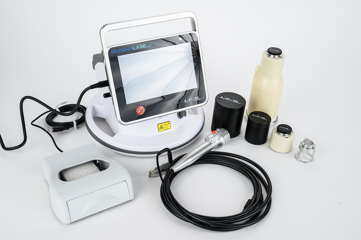
The Regenelase is a dual-laser system developed for musculoskeletal soft tissue. It operates at two distinct wavelengths, each serving a different biological role:
- 1550 nm erbium fiber laser (internal): Delivered via a 400-micron optical fiber threaded through an 18-gauge cannula — a standard injection needle — under image guidance. When the fiber tip contacts target cartilage, it delivers a precisely controlled burst of energy in approximately 50 seconds, creating a grid of microscopic zones of thermal coagulation within the tissue.
- 980 nm diode laser (external): Applied to the surface of the skin overlying the treated joint as a follow-up procedure. At this wavelength, the laser penetrates to deep tissue, delivering photobiomodulation (PBM) therapy that elevates tissue temperature, modulates local inflammation, reduces pain, and increases circulation to prepare the surrounding tissue for healing.
Together, they treat the joint from the inside out (fractional fiber laser creating channels in cartilage) and the outside in (photobiomodulation activating the surrounding tissue and preparing biologics). This dual approach addresses the joint environment comprehensively — not just the cartilage surface.
Three Mechanisms: What the Laser Is Actually Doing
When the 1550 nm laser contacts cartilage, it doesn’t ablate or remove tissue. It creates nanopores — microscopic channels in the extracellular matrix — through three simultaneous mechanisms confirmed by atomic force microscopy in animal studies:
The result is a structurally modified cartilage environment that is biologically active, receptive to healing factors, and capable of integration with newly delivered biologics. Histological analysis from animal studies has shown that this fractional coagulative treatment can generate hyaline-like cartilage in chondromalacia — the type of cartilage most similar to what was lost, not the fibrocartilage scar that most healing modalities produce.
Biologics can’t reach cartilage that hasn’t been prepared to receive them. The laser doesn’t just treat the tissue — it opens a door that was otherwise closed. — Dr. Trevor Turner, MD, RMSK · Founder, CartiNova & Pravida Health
The Biologics Layer: PRP, BMAC, and MFAT
Once the Regenelase laser has prepared the cartilage environment, the second layer of the CartiNova procedure delivers concentrated autologous biologics under image guidance — directly into the nanopore-modified tissue. Three primary biologic options are used based on the degree of degeneration and the patient’s BioProfile:
Neither element works as well in isolation. Biologics delivered without laser preparation are largely confined to the joint space and cannot penetrate avascular cartilage in meaningful concentrations. The laser alone activates cartilage healing mechanisms but has limited biological material to work with. Together, the laser creates the structural opportunity and the biologics supply the regenerative signals — a mechanism that doesn’t exist in any other single-modality treatment.
The CartiNova Procedure: Step by Step
Clinical Evidence: What the Data Shows
The Regenelase system has been evaluated in multiple clinical settings. Key findings from published and presented data:
- 90% of patients in clinical studies of knee osteoarthritis treated with the Regenelase fractional laser showed improved pain and function at both 6 and 12 months follow-up, with no side effects reported.
- MRI follow-up in patients treated at CartiNova has shown objective evidence of new cartilage growth within six months — the kind of structural change that “non-surgical” options have historically been unable to demonstrate.
- Atomic force microscopy in animal studies confirmed nanopore formation in laser-treated cartilage, with histological analysis showing hyaline-like cartilage regeneration — not the fibrocartilage scar that microfracture and other older techniques produce.
- Significant improvement across pain, emotional well-being, and general health scores at 12-month follow-up in knee OA patients, including resolution of bone marrow edema and improved cartilage thickness in some cases.
A real-world example that illustrates the clinical potential: a long-distance runner diagnosed with severe arthritis and told she would never run again underwent the CartiNova procedure and reported running 10 miles pain-free within three weeks of treatment. While outcomes vary by patient and degree of degeneration, this case reflects what the combination of laser-prepared cartilage and concentrated biologics can achieve in the right candidate.
The Regenelase evidence base, while growing, reflects a relatively recent technology. Large-scale randomized controlled trials are ongoing. As with any emerging regenerative procedure, we are honest with patients about what is established versus still accumulating. The FDA clearance, combined with the mechanistic rationale and clinical outcomes to date, form the basis of our use of this technology at Pravida Health.
Who Is a Candidate for CartiNova?
The CartiNova procedure is designed for patients who are in the gap between “take an anti-inflammatory and wait” and “you need surgery” — a gap that standard orthopedic care often ignores. Conditions we treat include:
- Knee cartilage lesions and osteoarthritis (mild to advanced, including cases where other physicians have recommended replacement)
- Lumbar disc degeneration and facet arthropathy
- Sports-related cartilage injuries in active patients who want to preserve long-term joint function
- Degenerative joint conditions in other major joints where image-guided delivery is feasible
- Post-surgical augmentation, to support healing after arthroscopic repair
Patients with active joint infection, severe coagulopathy, or truly end-stage bone-on-bone disease may not be appropriate candidates. An individual consultation including imaging review is the appropriate first step — candidacy depends on the specific anatomy, degree of degeneration, and individual BioProfile, not a blanket diagnosis.
Frequently Asked Questions
What is the Regenelase laser and how does it work on cartilage?
The Regenelase is a dual-laser system: a 1550 nm erbium fiber laser delivered through an 18-gauge needle that creates microscopic zones of thermal coagulation (nanopores) inside cartilage, and a 980 nm diode laser applied externally for photobiomodulation. The internal fractional laser works in approximately 50 seconds, creating a precision grid of channels that serve as biological scaffolding for healing factors. The external laser increases circulation and activates surrounding tissue. Together they treat the joint from the inside out and the outside in.
Is the CartiNova procedure FDA cleared?
Yes. The Regenelase system is FDA cleared (K233344). The 1550 nm laser is cleared for coagulating soft tissue including meniscus cartilage. The 980 nm diode laser is cleared for elevating tissue temperature to treat medical conditions including temporary relief of pain, muscle spasms, and increased local circulation. The procedure is performed under local anesthesia in an office setting — no general anesthesia or hospital admission required.
Why does cartilage fail to heal on its own?
Cartilage is avascular — it has no direct blood supply. Unlike bone or muscle, which recruit healing cells through the bloodstream after injury, cartilage relies on nutrient diffusion from synovial fluid. This process is slow, limited, and completely inadequate for meaningful repair of cartilage lesions or degenerative changes. Without a blood supply, the body’s normal inflammatory healing response simply cannot reach cartilage in meaningful concentrations.
How are biologics (PRP and BMAC) used in the CartiNova procedure?
After the Regenelase laser creates nanopores in the cartilage, concentrated biologics — PRP, BMAC, or MFAT — are delivered directly to the prepared tissue under image guidance. The nanopores provide channels that allow growth factors and regenerative cells to penetrate the cartilage matrix, rather than remaining on the surface or in the joint fluid where their access to cartilage is limited.
What results has the Regenelase laser shown in clinical studies?
Clinical studies of the Regenelase system for knee osteoarthritis have shown that 90% of patients demonstrated improvement in pain and function at both 6 and 12 months follow-up, with no side effects reported. Follow-up MRI in some patients has shown objective evidence of new cartilage growth within six months. Animal studies using atomic force microscopy confirmed nanopore formation in laser-treated cartilage, with histological analysis showing hyaline-like cartilage regeneration.
Who is a candidate for the CartiNova procedure?
Ideal candidates are patients with cartilage lesions, knee osteoarthritis (mild to advanced), lumbar disc degeneration, sports-related cartilage injuries, or degenerative joint conditions seeking a non-surgical alternative. The CartiNova BioProfile — combining whole genome sequencing, advanced biomarkers, imaging, and AI analysis — identifies the precise drivers of joint degeneration and tailors the procedure to each patient. An individual consultation with imaging review is the appropriate first step.
What is the recovery time after a CartiNova procedure?
Recovery is significantly shorter than surgical alternatives. Patients are typically ambulatory immediately after the procedure. The biologics require a short period of reduced loading — typically 24 to 72 hours of modified activity — before progressive rehabilitation begins. The full regenerative process occurs over weeks to months, with OxeFit AI-guided analytics tracking functional improvement throughout recovery.
Ready to Find Out If CartiNova Is Right for You?
Candidacy depends on your specific anatomy, imaging findings, and degree of joint degeneration — not a blanket diagnosis. A consultation with Dr. Turner includes a review of your imaging, a frank discussion of what the evidence supports for your specific condition, and a clear explanation of which biologics and protocols are appropriate for your BioProfile.
Schedule a Consultation Learn About CartiNovaLearn more at cartinova.com · Existing patient? Patient Portal